
Dear Women's Imaging Insider,
The New England Journal of Medicine today published yet another study that calls into question the efficacy of screening mammography, further fueling a controversy that shows no signs of resolution.
In the paper, a group led by Dr. H. Gilbert Welch of Dartmouth College posits that the reduction in breast cancer mortality after the advent of screening mammography is mostly a result of improved therapy, not earlier detection.
Of course, critics' response to this conclusion has been swift. Check out our coverage of the study -- and one prominent breast radiologist's response.
Then take a look at our Insider Exclusive, which outlines the difficulty of providing breast cancer care in Afghanistan during wartime. A U.S. Air Force surgeon described how he and colleagues diagnosed, treated, and tracked Afghan women with breast cancer at Craig Joint Theater Hospital at Bagram Airfield, about 30 miles north of Kabul. Not surprisingly, they encountered some challenges.
When you've read our Insider feature, check out what else is going on in the Women's Imaging Community:
- Find out how contrast can boost the diagnostic power of breast ultrasound.
- Read what Yale University researchers have to say about the benefits of digital breast tomosynthesis in the diagnostic setting.
- Discover what a coalition of patient groups and breast cancer experts are doing to protect annual screening mammography coverage.
- Find out how PET can help predict outcomes in vulvar cancer patients.
- Read how Twitter can offer insights into women's perspectives on mammography screening.
If you have a comment, report, or article idea to share about any aspect of women's imaging, please contact me.

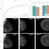






![A normal mammogram confirmed by three-year radiologic follow-up illustrates reader-marked regions of interest (ROIs) during (A) unaided (round 1) and (B) artificial intelligence (AI)–assisted (round 2) reading. Each colored dot represents an ROI for recall by a human reader. Readers could mark more than one ROI per case, represented by multiple dots of the same color. During AI-assisted reading, the AI system displayed three visible prompts: two with suspicion of malignancy scores of 35% (left mediolateral oblique [L MLO] and craniocaudal [L CC]) and one with a suspicion of malignancy score of 10% (right craniocaudal [R CC]), shown as polygonal overlays. Without AI, six of 10 readers (60%) marked a false-positive ROI. With AI assistance, this fell to two of 10 (20%). R MLO = right mediolateral oblique.](https://img.auntminnie.com/mindful/smg/workspaces/default/uploads/2026/07/2026-07-14-radiology-mammogram-ai-auto-bias.H0bYO8QlWs.jpg?auto=format%2Ccompress&dpr=2&fit=crop&h=167&q=70&w=250)


