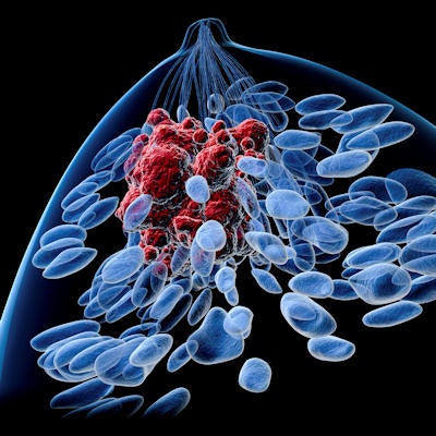
Contrast-enhanced ultrasound (CEUS) can be an effective method for the differential diagnosis of benign and malignant breast lesions, offering improvements in diagnostic accuracy over conventional breast ultrasound, according to research published in the October issue of the Journal of Ultrasound in Medicine.
In a meta-analysis incorporating nearly 30 studies and more than 2,000 patients, a team from Sichuan University in Chengdu, China, found that contrast-enhanced ultrasound offered a combined sensitivity of 88% and specificity of 80%. What's more, diagnostic performance also improved with the advent of second-generation contrast agents.
 Dr. Xuelei Ma from Sichuan University.
"Contrast-enhanced sonography could be developed as a promising and effective tool for distinguishing benign from malignant breast lesions, which may be a valuable supplement to fine-needle aspiration [biopsy]," first author Dr. Xuelei Ma told AuntMinnie.com. "Meanwhile, in terms of contrast-enhanced breast sonography, the second-generation contrast agents might increase the diagnostic accuracy, and the harmonic mode might be more suitable for performance."
Improving accuracy?
Breast ultrasound has been shown to have relatively high sensitivity of approximately 90% and specificity of around 40%, according to the researchers. CEUS has been suggested as a way to improve the modality's diagnostic accuracy, but relevant studies have shown sensitivity ranging from 50% to 100% and specificity ranging from 37% to 100%.
"Meanwhile, the overall analysis of different types of contrast agents and contrast modes has not been assessed systematically," Ma said. "Therefore, we carried out this study to investigate the performance of contrast-enhanced sonography in the differential diagnosis of benign and malignant breast lesions."
The researchers searched PubMed and Embase for all relevant studies performed before January 2015. They also manually reviewed references of related articles (J Ultrasound Med, October 2016, Vol. 35:10, pp. 2,095-2,102).
Clinical studies were included in the meta-analysis if they met the following criteria:
- They focused on the diagnostic value of contrast-enhanced sonography in breast cancer.
- Histopathologic findings were used as the reference standard for lesion diagnosis.
- They offered sufficient data to construct a table corresponding to true-positive, true-negative, false-positive, and false-negative rates.
- The sample size must have been more than 10 patients and duplicate results needed to be eliminated.
The researchers did not include letters, reviews, editorials, and case reports. In cases where a study involved the same author or authors from the same medical center and had similar data and patient design, the researchers selected the article with the larger sample size. A total of 29 eligible studies satisfied the inclusion criteria and included an aggregate of 2,234 patients and 2,929 lesions.
Next, the team used the true-positive, false-positive, false-negative, and true-negative rates from all of the studies to produce a summary of sensitivity, specificity, positive likelihood ratios, negative likelihood ratios, and diagnostic odds ratio. They constructed a receiver operator characteristics (ROC) curve to summarize the true-positive and false-positive rates. They also assessed the quality of the studies using version 2 of the Quality Assessment of Diagnostic Accuracy Studies (QUADAS-2) method.
CEUS showed high sensitivity and specificity:
- Sensitivity: 88%
- Specificity: 80%
- Pooled diagnostic odds ratio: 30.35
- Area under the curve: 0.9115
Newer agents perform better
In further analysis, the researchers also compared the results of studies that used first-generation ultrasound contrast agents such as Levovist (Schering) and second-generation agents such as SonoVue (Bracco Diagnostics). In addition, they assessed the type of contrast mode used in each study.
"We found that the second-generation agent group had relatively higher sensitivity and specificity compared with the first-generation group, and the diagnostic performance was significantly higher in the harmonic mode in comparison with the power/color Doppler mode," Ma said.
| Performance by contrast media type | ||||
| Sensitivity | Specificity | Diagnostic odds ratio | Area under the curve | |
| 1st-generation contrast agents | 86% | 77% | 26.3 | 0.81 |
| 2nd-generation contrast agents | 90% | 85% | 47.2 | 0.94 |
| Performance by contrast mode | ||||
| Sensitivity | Specificity | Diagnostic odds ratio | Area under the curve | |
| Power/color Doppler | 85% | 69% | 18.7 | 0.88 |
| Harmonic | 90% | 87% | 51.6 | 0.94 |
"Our study suggests that contrast-enhanced sonography is a promising and effective method for differential diagnosis of benign and malignant breast lesions," the authors wrote. "It may be a valuable supplement to fine-needle aspiration [biopsy], which is still needed in cases of Breast Imaging Reporting and Data System category 3 and 4 lesions."
Further studies are still required to confirm their findings, according to the group. The researchers also plan to investigate the performance of CEUS in the differential diagnosis of benign and malignant lesions other than breast cancer.