Fujifilm Europe is highlighting several new offerings at this year's ECR 2015 meeting in Vienna.
The company's Medical Systems Division has just released its FDR D-EVOII digital radiography (DR) panel, which includes a shell rib magnesium frame that reduces its weight by 20% compared to the company's FDR D-EVO flat-panel detectors, according to Fujifilm. The panel is available in 35 x 43-cm and 43 x 43-cm cesium iodide (CsI) and gadolinium oxysulfide (GOS) models. A 24 x 30-cm CsI model will be released in the coming months, the company said.
As well, the company has released upgrades to its FDR Amulet Innovality digital mammography device. New features include the option to execute biopsy targeting using tomosynthesis images, a work-in-progress improved version of Amulet Innovality operator console software for use with the tomosynthesis biopsy function, a new patented shifting paddle, and a new optional height-adjustable operator console, according to Fujifilm Europe.
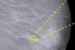
Also in its ECR booth, Fujifilm will introduce a new image processing tool called Virtual Grid, which maps the effect of scatter upon each image before automatically adapting the processing to replicate grid use and reduce degradation of image quality.
Finally, Fujifilm Europe will showcase its Synapse portfolio, including Synapse VNA, Synapse 3D, Synapse Clinical Workflow Manager, and Synapse ERm, the company said.

![A normal mammogram confirmed by three-year radiologic follow-up illustrates reader-marked regions of interest (ROIs) during (A) unaided (round 1) and (B) artificial intelligence (AI)–assisted (round 2) reading. Each colored dot represents an ROI for recall by a human reader. Readers could mark more than one ROI per case, represented by multiple dots of the same color. During AI-assisted reading, the AI system displayed three visible prompts: two with suspicion of malignancy scores of 35% (left mediolateral oblique [L MLO] and craniocaudal [L CC]) and one with a suspicion of malignancy score of 10% (right craniocaudal [R CC]), shown as polygonal overlays. Without AI, six of 10 readers (60%) marked a false-positive ROI. With AI assistance, this fell to two of 10 (20%). R MLO = right mediolateral oblique.](https://img.auntminnie.com/mindful/smg/workspaces/default/uploads/2026/07/2026-07-14-radiology-mammogram-ai-auto-bias.H0bYO8QlWs.jpg?auto=format%2Ccompress&fit=crop&h=100&q=70&w=100)


![A normal mammogram confirmed by three-year radiologic follow-up illustrates reader-marked regions of interest (ROIs) during (A) unaided (round 1) and (B) artificial intelligence (AI)–assisted (round 2) reading. Each colored dot represents an ROI for recall by a human reader. Readers could mark more than one ROI per case, represented by multiple dots of the same color. During AI-assisted reading, the AI system displayed three visible prompts: two with suspicion of malignancy scores of 35% (left mediolateral oblique [L MLO] and craniocaudal [L CC]) and one with a suspicion of malignancy score of 10% (right craniocaudal [R CC]), shown as polygonal overlays. Without AI, six of 10 readers (60%) marked a false-positive ROI. With AI assistance, this fell to two of 10 (20%). R MLO = right mediolateral oblique.](https://img.auntminnie.com/mindful/smg/workspaces/default/uploads/2026/07/2026-07-14-radiology-mammogram-ai-auto-bias.H0bYO8QlWs.jpg?auto=format%2Ccompress&fit=crop&h=112&q=70&w=112)