Dear AuntMinnie Member,
Our virtual conference, PACS in an Age of Change, starts tomorrow, November 17 -- have you registered yet?
If not, there's still time. By registering, you'll be able to attend six presentations on PACS by some of the most important thought leaders in radiology -- speakers like Katherine Andriole, PhD, of Brigham and Women's Hospital in Boston. Dr. Andriole will speak at 4 p.m. EST on informatics tools to improve radiology reporting; specifically, she'll share how her facility improved its critical results reporting.
To view a list of the other speakers and to get more information about the conference, just go to pacschange.auntminnie.com. See you Wednesday!
PEM and MBI for breast imaging
Nuclear medicine-based technologies for breast imaging are grabbing headlines in medical imaging this week. Last week, a new study was published in Radiology that compared positron emission mammography (PEM) to breast MRI in terms of each technology's impact on surgical management of women with ipsilateral breast cancer.
The study found that PEM's strong suit was finding benign breast lesions, which could reduce unnecessary biopsies. But how well did PEM perform in other areas? Find out by clicking here.
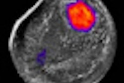
Meanwhile, researchers from the Mayo Clinic in Rochester, MN, examined another nuclear breast imaging modality, molecular breast imaging (MBI), which is based on a gamma camera with dual-head digital detectors.
The study compared MBI to conventional mammography in women with dense breasts -- historically a weak point for x-ray-based imaging. The group found that MBI performed far better than mammography in this patient population -- find out how much by clicking here, or visit our Women's Imaging Digital Community at women.auntminnie.com.

![A normal mammogram confirmed by three-year radiologic follow-up illustrates reader-marked regions of interest (ROIs) during (A) unaided (round 1) and (B) artificial intelligence (AI)–assisted (round 2) reading. Each colored dot represents an ROI for recall by a human reader. Readers could mark more than one ROI per case, represented by multiple dots of the same color. During AI-assisted reading, the AI system displayed three visible prompts: two with suspicion of malignancy scores of 35% (left mediolateral oblique [L MLO] and craniocaudal [L CC]) and one with a suspicion of malignancy score of 10% (right craniocaudal [R CC]), shown as polygonal overlays. Without AI, six of 10 readers (60%) marked a false-positive ROI. With AI assistance, this fell to two of 10 (20%). R MLO = right mediolateral oblique.](https://img.auntminnie.com/mindful/smg/workspaces/default/uploads/2026/07/2026-07-14-radiology-mammogram-ai-auto-bias.H0bYO8QlWs.jpg?auto=format%2Ccompress&fit=crop&h=100&q=70&w=100)


![A normal mammogram confirmed by three-year radiologic follow-up illustrates reader-marked regions of interest (ROIs) during (A) unaided (round 1) and (B) artificial intelligence (AI)–assisted (round 2) reading. Each colored dot represents an ROI for recall by a human reader. Readers could mark more than one ROI per case, represented by multiple dots of the same color. During AI-assisted reading, the AI system displayed three visible prompts: two with suspicion of malignancy scores of 35% (left mediolateral oblique [L MLO] and craniocaudal [L CC]) and one with a suspicion of malignancy score of 10% (right craniocaudal [R CC]), shown as polygonal overlays. Without AI, six of 10 readers (60%) marked a false-positive ROI. With AI assistance, this fell to two of 10 (20%). R MLO = right mediolateral oblique.](https://img.auntminnie.com/mindful/smg/workspaces/default/uploads/2026/07/2026-07-14-radiology-mammogram-ai-auto-bias.H0bYO8QlWs.jpg?auto=format%2Ccompress&fit=crop&h=112&q=70&w=112)