Wednesday, December 1 | 11:50 a.m.-12:00 p.m. | SSK01-09 | Room E450A
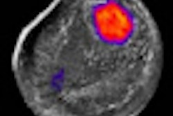
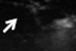
Examining excised breast tissue with high-resolution ultrasound elastography could help surgeons confirm tumor-free margins with greater accuracy than specimen mammography, according to this Wednesday morning presentation by German researchers.Presenter Reinhard Kubale, MD, PhD, will discuss how his team used high-resolution ultrasound and specimen mammography to examine specimens resected from 55 patients with biopsy-confirmed tumors. High-resolution ultrasound was performed during operations using a 17- and 18-MHz scan head with Hannafy lens (Acuson S2000, Siemens Healthcare, Erlangen, Germany).
Re-excision was performed for patients with close (< 5 mm) or positive margins, and tumor-free distance was compared to histology.
Kubale and colleagues found good visibility of tumors and margins in 51 of 55 cases. There was an 89% correlation between histological and high-resolution ultrasound measurements of tumor-free margin. Re-excision was performed in 31 cases, with intraoperative re-excision correct in 26 of 31 cases.
High-resolution ultrasound was superior to specimen mammography in detecting resected tumor and tumor-free border, and it reduced the reoperation rate from 12% to 5%, the authors concluded.

![A normal mammogram confirmed by three-year radiologic follow-up illustrates reader-marked regions of interest (ROIs) during (A) unaided (round 1) and (B) artificial intelligence (AI)–assisted (round 2) reading. Each colored dot represents an ROI for recall by a human reader. Readers could mark more than one ROI per case, represented by multiple dots of the same color. During AI-assisted reading, the AI system displayed three visible prompts: two with suspicion of malignancy scores of 35% (left mediolateral oblique [L MLO] and craniocaudal [L CC]) and one with a suspicion of malignancy score of 10% (right craniocaudal [R CC]), shown as polygonal overlays. Without AI, six of 10 readers (60%) marked a false-positive ROI. With AI assistance, this fell to two of 10 (20%). R MLO = right mediolateral oblique.](https://img.auntminnie.com/mindful/smg/workspaces/default/uploads/2026/07/2026-07-14-radiology-mammogram-ai-auto-bias.H0bYO8QlWs.jpg?auto=format%2Ccompress&fit=crop&h=100&q=70&w=100)


![A normal mammogram confirmed by three-year radiologic follow-up illustrates reader-marked regions of interest (ROIs) during (A) unaided (round 1) and (B) artificial intelligence (AI)–assisted (round 2) reading. Each colored dot represents an ROI for recall by a human reader. Readers could mark more than one ROI per case, represented by multiple dots of the same color. During AI-assisted reading, the AI system displayed three visible prompts: two with suspicion of malignancy scores of 35% (left mediolateral oblique [L MLO] and craniocaudal [L CC]) and one with a suspicion of malignancy score of 10% (right craniocaudal [R CC]), shown as polygonal overlays. Without AI, six of 10 readers (60%) marked a false-positive ROI. With AI assistance, this fell to two of 10 (20%). R MLO = right mediolateral oblique.](https://img.auntminnie.com/mindful/smg/workspaces/default/uploads/2026/07/2026-07-14-radiology-mammogram-ai-auto-bias.H0bYO8QlWs.jpg?auto=format%2Ccompress&fit=crop&h=112&q=70&w=112)