Wednesday, December 1 | 8:35 a.m.-8:45 a.m. | VB41-01 | Arie Crown Theater
In this paper presentation, researchers will discuss study results showing contrast-enhanced spectral mammography's (CESM) potential to increase the performance and sensitivity of standard mammography and breast ultrasound.Clarisse Dromain, MD, and colleagues from Institut Gustave-Roussy in Villejuif, France, will present findings from a study they conducted to evaluate the clinical performance of this technology as an adjunct to mammography and ultrasound.
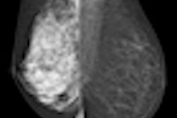
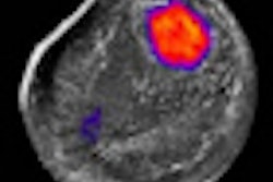
CESM uses images acquired at different x-ray energy levels to identify breast tumors. One image is taken in the conventional mammography range of 26-32 kVp and provides morphological information; another is taken using voltages in the 45-49 kVp range, which captures the diffusion of a contrast agent within the breast. The two images are then combined into one.
The study included 120 women recalled for diagnostic workup who underwent mammography, ultrasound, and CESM exams. The team used a Senographe DS system (GE Healthcare, Chalfont St. Giles, U.K.) that had been modified to perform the CESM exams. Each woman's actual condition was checked with histology, cytology, or a one-year follow-up.
The team found 150 lesions, of which 86 were malignant and 64 were benign. The clinical performance in interpreting the lesions was measured for seven breast radiologists who were blinded to the results. For conventional mammography plus ultrasound, the readers turned in an area under the receiver operator characteristics (ROC) curve of 0.83, a number that increased to 0.88 for CESM with ultrasound.
This led Dromain's team to conclude that adding the technology to conventional modalities such as mammography and ultrasound could be a valuable technique for diagnostic patients.


![A normal mammogram confirmed by three-year radiologic follow-up illustrates reader-marked regions of interest (ROIs) during (A) unaided (round 1) and (B) artificial intelligence (AI)–assisted (round 2) reading. Each colored dot represents an ROI for recall by a human reader. Readers could mark more than one ROI per case, represented by multiple dots of the same color. During AI-assisted reading, the AI system displayed three visible prompts: two with suspicion of malignancy scores of 35% (left mediolateral oblique [L MLO] and craniocaudal [L CC]) and one with a suspicion of malignancy score of 10% (right craniocaudal [R CC]), shown as polygonal overlays. Without AI, six of 10 readers (60%) marked a false-positive ROI. With AI assistance, this fell to two of 10 (20%). R MLO = right mediolateral oblique.](https://img.auntminnie.com/mindful/smg/workspaces/default/uploads/2026/07/2026-07-14-radiology-mammogram-ai-auto-bias.H0bYO8QlWs.jpg?auto=format%2Ccompress&fit=crop&h=100&q=70&w=100)


![A normal mammogram confirmed by three-year radiologic follow-up illustrates reader-marked regions of interest (ROIs) during (A) unaided (round 1) and (B) artificial intelligence (AI)–assisted (round 2) reading. Each colored dot represents an ROI for recall by a human reader. Readers could mark more than one ROI per case, represented by multiple dots of the same color. During AI-assisted reading, the AI system displayed three visible prompts: two with suspicion of malignancy scores of 35% (left mediolateral oblique [L MLO] and craniocaudal [L CC]) and one with a suspicion of malignancy score of 10% (right craniocaudal [R CC]), shown as polygonal overlays. Without AI, six of 10 readers (60%) marked a false-positive ROI. With AI assistance, this fell to two of 10 (20%). R MLO = right mediolateral oblique.](https://img.auntminnie.com/mindful/smg/workspaces/default/uploads/2026/07/2026-07-14-radiology-mammogram-ai-auto-bias.H0bYO8QlWs.jpg?auto=format%2Ccompress&fit=crop&h=112&q=70&w=112)