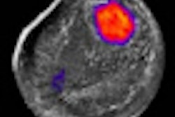
Researchers in the U.K. are preparing to launch a 1.6 million pound ($2.6 million U.S.) trial to determine the efficacy of digital breast tomosynthesis (DBT) to screen for breast cancer.
Approximately 7,000 women in Aberdeen, Glasgow, Manchester, London, and Guildford will be invited to participate in the three-year study.
Professor Fiona Gilbert, head of imaging at the University of Aberdeen, is the chief investigator of the study, which is funded by a grant from the National Institute for Health Research (NIHR) Health Technology Assessment program.
Women who are recalled after an abnormal screening mammogram will be invited to participate in the trial, which will begin recruiting volunteers early next year. Trial participants will receive a standard mammogram and a DBT examination. The 2D and 3D images will be collected and reviewed independently by radiologists at another facility.
The research team will then compare the number of cancer cases detected by each imaging technique, the number of false positives, and the relative effectiveness of DBT in women with dense-tissue breasts, which can be a cancer risk.
Related Reading
Norwegian researcher launches DBT study, October 4, 2010
FDA panel gives nod to Hologic tomosynthesis PMA, September 24, 2010
Study offers comparison of conebeam breast CT to mammo, July 27, 2010
CARS report: Novel conebeam CT cuts breast dose, improves accuracy, June 29, 2009
ECR delivers new findings on digital breast tomosynthesis, March 7, 2009
Copyright © 2010 AuntMinnie.com

![A normal mammogram confirmed by three-year radiologic follow-up illustrates reader-marked regions of interest (ROIs) during (A) unaided (round 1) and (B) artificial intelligence (AI)–assisted (round 2) reading. Each colored dot represents an ROI for recall by a human reader. Readers could mark more than one ROI per case, represented by multiple dots of the same color. During AI-assisted reading, the AI system displayed three visible prompts: two with suspicion of malignancy scores of 35% (left mediolateral oblique [L MLO] and craniocaudal [L CC]) and one with a suspicion of malignancy score of 10% (right craniocaudal [R CC]), shown as polygonal overlays. Without AI, six of 10 readers (60%) marked a false-positive ROI. With AI assistance, this fell to two of 10 (20%). R MLO = right mediolateral oblique.](https://img.auntminnie.com/mindful/smg/workspaces/default/uploads/2026/07/2026-07-14-radiology-mammogram-ai-auto-bias.H0bYO8QlWs.jpg?auto=format%2Ccompress&fit=crop&h=100&q=70&w=100)


![A normal mammogram confirmed by three-year radiologic follow-up illustrates reader-marked regions of interest (ROIs) during (A) unaided (round 1) and (B) artificial intelligence (AI)–assisted (round 2) reading. Each colored dot represents an ROI for recall by a human reader. Readers could mark more than one ROI per case, represented by multiple dots of the same color. During AI-assisted reading, the AI system displayed three visible prompts: two with suspicion of malignancy scores of 35% (left mediolateral oblique [L MLO] and craniocaudal [L CC]) and one with a suspicion of malignancy score of 10% (right craniocaudal [R CC]), shown as polygonal overlays. Without AI, six of 10 readers (60%) marked a false-positive ROI. With AI assistance, this fell to two of 10 (20%). R MLO = right mediolateral oblique.](https://img.auntminnie.com/mindful/smg/workspaces/default/uploads/2026/07/2026-07-14-radiology-mammogram-ai-auto-bias.H0bYO8QlWs.jpg?auto=format%2Ccompress&fit=crop&h=112&q=70&w=112)