Dear AuntMinnie Member,
In 2006, the U.S. Food and Drug Administration proposed to relax the rules under which it approves full-field digital mammography (FFDM) systems. Three years later, mammography vendors are still waiting.
The FDA's proposal was to reclassify FFDM systems so they would be reviewed under the 510(k) process, which typically takes 90 days, rather than the premarket approval process, which requires clinical trials to be performed and can take up to a year or more.
So what gives? The FDA isn't talking, but some mammography advocates and device vendors are grumbling that the agency's delay is making advanced breast imaging technology that's available in the rest of the world inaccessible to U.S. patients. Read associate editor Kate Madden Yee's article by clicking here.
In other news, is it possible for tumors found on mammography screening to "spontaneously regress," ultimately causing no harm to the patient? This controversial theory was revived last week in a symposium on breast cancer diagnosis and treatment. Find out what the fuss was all about by clicking here, or visit our Women's Imaging Digital Community at women.auntminnie.com.
VC for flat polyps
In other news, flat polyps have historically vexed proponents of virtual colonoscopy, but new research presented at this month's American College of Radiology Imaging Network (ACRIN) fall meeting indicates that they may not be so tough to find after all.
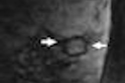
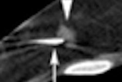
Researchers from the Mayo Clinic in Rochester, MN, analyzed data from the National CT Colonography Trial (ACRIN 6664) and found that flat polyps actually are visible on VC exams -- and picking them up might just be a matter of proper training.
Read all about it by clicking here for an article by international editor Eric Barnes in our Virtual Colonoscopy Digital Community, or go to vc.auntminnie.com.