
Breast MRI can help doctors put equivocal mammographic findings to rest, but it should be used carefully because of the potential for false positives, according to a new study in the October issue of the American Journal of Roentgenology.
Researchers at New York University (NYU) School of Medicine in New York City determined that breast MRI was particularly helpful when the ambiguous mammographic finding was breast asymmetry or architectural distortion -- helpful, but not always necessary. Dr. Linda Moy and colleagues cautioned that when using MRI to resolve these findings, strict selection criteria should be observed because of the frequency of incidental and false-positive lesions seen with the modality (AJR, October 2009, Vol. 193:4, pp. 986-993).
"More clinicians and radiologists are ordering breast MRI to further investigate a mammogram finding because patients don't want to undergo a biopsy, because the doctor wants to be extra vigilant in case of litigation, or because the abnormality has not been thoroughly worked up on mammography and ultrasound," Moy told AuntMinnie.com. "Breast MRI adds a lot of information, and it may resolve the question. But because the exam may also find incidental lesions, it should be used with care."
Of 80,526 mammograms conducted between 1999 and 2005 at the Breast Imaging Center at the NYU Cancer Institute, 115 breast MRI exams were performed to follow up inconclusive findings. Moy's team reviewed these 115 exams and found that 48 (41.8%) were from high-risk women, 12 (10.4%) were from BRCA-1 or BRCA-2 carriers, 20 (17.4%) were from women with a personal history of breast cancer, and 16 (13.9%) were from patients with a previous biopsy finding of a high-risk lesion.
The mammographic abnormalities that prompted breast MRI included asymmetry without microcalcifications (98 of 115, or 85.2%), architectural distortion (12, or 10.4%), and change in appearance at the site of a previously benign biopsy (five, or 4.3%).
 |
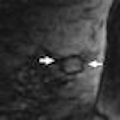
| The following images are of a 46-year-old woman with one-view asymmetry in right breast. Above, craniocaudal projection mammogram of both breasts shows one-view asymmetry (arrow) in medial aspect of right breast. All images courtesy of the American Roentgen Ray Society. |
 |
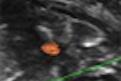
| Contrast-enhanced fat-suppressed T1-weighted sagittal MR image of right breast shows mildly enhancing mass (arrows) that corresponds to asymmetry. |
 |
| True lateral (left) and craniocaudal (right) projection mammograms of right breast after needle localization show asymmetry has been localized on two views. Final pathologic finding was stromal fibrosis. |
No further suspicious findings were discovered in 100 of the 115 cases, which were found to be stable in mammography or MRI follow-up after a mean of 34 months. Of the 15 malignancies, nine were surgically removed and yielded benign findings; the remaining six proved malignant after surgery.
The study found 18 incidental lesions out of the 115 breast MRI exams. None proved malignant and, in fact, they just increased the cost of evaluating the first inconclusive mammographic findings, according to Moy.
"For example, we excluded palpable lumps and calcifications from this study," Moy said. "Surgical removal of the lump and breast biopsy of the calcification are more definitive ways to resolve the ambiguity than breast MRI."
Do BI-RADS characteristics point to malignancy?
Also in the October AJR issue, researchers at the University of Washington Medical Center in Seattle found that certain combinations of BI-RADS lesion descriptors can predict the probability of malignancy for breast MRI masses (pp. 994-1000).
The idea that particular BI-RADS categories can point to malignancy has been true in mammography, but it has not been explicitly shown for MRI, according to lead author Dr. Robert Gutierrez.
"[Our goal in this study was to help] improve the diagnostic accuracy of breast MRI," he told AuntMinnie.com. "The ultimate goal is to develop an algorithm based on BI-RADS categories that radiologists can use when they sit down to read these exams."
The study included 196 women with 258 suspicious lesions that were collected from breast MRI exams performed between January 2003 and June 2005. Among all lesions, 147 (57%) were 1 cm or greater and 111 (43%) were less than 1 cm. The 258 lesions included 123 (48%) masses, 95 (37%) nonmasslike enhancements, and 40 (16%) focus or foci.
Gutierrez's team found that a particular combination of BI-RADS characteristics tended to signify malignancy in breast MRI.
"If a mass is greater than 1 cm, is heterogeneously enhancing, and has irregular or spiculated margins, we found a 68% probability that it will be malignant," he said.
 |
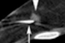
| Contrast-enhanced T1-weighted sagittal image in 62-year-old woman shows 24-mm mass with morphologic features common to malignant masses in this study: large size, irregular shape, irregular margins, and heterogeneous enhancement. |
The results indicate that breast MRI's diagnostic accuracy needs refining, Gutierrez said.
"Most radiologists don't have a problem deciding whether a mass needs to be biopsied. The problem comes in when they're dealing with equivocal lesions -- those in the gray zone," he said.
By Kate Madden Yee
AuntMinnie.com staff writer
October 6, 2009
Related Reading
Patient age, clinical indication need to be considered in breast MRI, September 18, 2009
CAD enhances breast MRI specificity, September 17, 2009
Breast lesion kinetic curves inconsistent across MRI systems, August 25, 2009
Kinetic information identifies MRI-detected breast tumors, August 19, 2009
Study: MRI may cause more harm than good in breast cancer, August 13, 2009
Copyright © 2009 AuntMinnie.com

![A normal mammogram confirmed by three-year radiologic follow-up illustrates reader-marked regions of interest (ROIs) during (A) unaided (round 1) and (B) artificial intelligence (AI)–assisted (round 2) reading. Each colored dot represents an ROI for recall by a human reader. Readers could mark more than one ROI per case, represented by multiple dots of the same color. During AI-assisted reading, the AI system displayed three visible prompts: two with suspicion of malignancy scores of 35% (left mediolateral oblique [L MLO] and craniocaudal [L CC]) and one with a suspicion of malignancy score of 10% (right craniocaudal [R CC]), shown as polygonal overlays. Without AI, six of 10 readers (60%) marked a false-positive ROI. With AI assistance, this fell to two of 10 (20%). R MLO = right mediolateral oblique.](https://img.auntminnie.com/mindful/smg/workspaces/default/uploads/2026/07/2026-07-14-radiology-mammogram-ai-auto-bias.H0bYO8QlWs.jpg?auto=format%2Ccompress&fit=crop&h=100&q=70&w=100)


![A normal mammogram confirmed by three-year radiologic follow-up illustrates reader-marked regions of interest (ROIs) during (A) unaided (round 1) and (B) artificial intelligence (AI)–assisted (round 2) reading. Each colored dot represents an ROI for recall by a human reader. Readers could mark more than one ROI per case, represented by multiple dots of the same color. During AI-assisted reading, the AI system displayed three visible prompts: two with suspicion of malignancy scores of 35% (left mediolateral oblique [L MLO] and craniocaudal [L CC]) and one with a suspicion of malignancy score of 10% (right craniocaudal [R CC]), shown as polygonal overlays. Without AI, six of 10 readers (60%) marked a false-positive ROI. With AI assistance, this fell to two of 10 (20%). R MLO = right mediolateral oblique.](https://img.auntminnie.com/mindful/smg/workspaces/default/uploads/2026/07/2026-07-14-radiology-mammogram-ai-auto-bias.H0bYO8QlWs.jpg?auto=format%2Ccompress&dpr=2&fit=crop&h=167&q=70&w=250)


