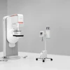
A new breast imaging technique could offer 3-D mammograms as an alternative to conventional 2-D digital mammography. At last month's RSNA meeting, a researcher from Wake Forest University presented results from an emerging technology known as tuned-aperture computed tomography (TACT).
TACT collects 2-D slices of the breast from different angles. The slices are then reconstructed into a 3-D image of the breast, which can be reviewed on a workstation. The technique was developed by Finnish mammography firm Instrumentarium.
At the RSNA conference, Dr. Richard Webber of the departments of dentistry and radiology at Wake Forest in Winston-Salem, NC, discussed his work with TACT.
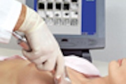
Most practical noninvasive methods for generating 3-D images require sufficient breast stabilization during data acquisition. However, unpredictable movement is inevitable.
TACT creates relatively isotropic voxels through the nonlinear biorthogonal merging of independent tomographic representations, according to Webber.
"The basic idea underlying the research is to simply merge tomographic data produced from asymmetrical projection geometries and improve sampling," Webber said. "It involves retrospective analysis of any number of 2-D projections produced from any number of unknown projection geometries."
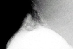
For this test, Webber used a cube-shaped specimen containing human breast tissue with reference points attached to each corner. The specimen was irradiated from five angles and from two orthogonal orientations for a total of 10 projections.
The resulting data was processed using TACT to produce two independent laminographic series that were derived from the same tissues, but oriented orthogonally.
"In order to merge this independent series of projection data, one has to correct for cone-beam artifacts," Webber said.

A flow chart of the TACT experiment
Relationships derived from respective projections of the attached reference points were used to make these corrections. A matrix-rotation program was applied to the data underlying the affine-corrected 3-D representations, effectively flipping it 90º. By combining the two representations, Webber said he was able to assign more accurate estimates of isotropic volume elements in the resulting 3-D reconstructions. The technique was used on data from a conventional stereotactic mammogram.
"The specimen was frozen breast tissue known to contain details of diagnostic images such as calcified blood vessels and microcalcifications," Webber said. "Each set of projections was analyzed. Then they were synthesized into a stack. This rotation of the stack at orientated angles allowed us to measure voxel-by-voxel projections. The point is that you can see details that are remarkably blur-free for tomosynthetic construction models."
The ability to acquire data in short bursts without restricting object motion between bursts may offer an alternative to conventional 2-D digital mammography, as well as intrinsically anisotropic 3-D imaging, Webber concluded.
By Shalmali PalAuntMinnie.com staff writer
December 28, 2000
Related Reading
Instrumentarium to debut Diamond system in step towards full-field digital mammo, April 7, 2000
Click here to post your comments about this story. Please include the headline of the article in your message.
Copyright © 2000 AuntMinnie.com