A combined MRI approach can characterize architectural distortion (AD) changes on mammograms, suggest findings published August 15 in Magnetic Resonance Imaging.
A team led by Deshuo Dong from the First Affiliated Hospital of Dalian Medical University in Liaoning, China, found that combining intravoxel incoherent motion (IVIM) with dynamic contrast-enhanced (DCE-MRI) achieved perfect sensitivity and negative predictive value in a pilot study. The combination also showed high efficiency in diagnosing architectural distortions.
“IVIM combined with DCE-MRI is a reliable predictive tool for the classification of architectural distortions changes with high diagnostic efficacy,” Dong and co-authors wrote.
Previous studies have shown that DCE-MRI combined with apparent diffusion coefficient is more reliable than mammography in distinguishing breast lesions that appear as primary architectural distortions on mammograms.
IVIM is an MRI technique that provides information on both the diffusion of water and the perfusion of blood within a tissue. It separates diffusion and blood flow to the MRI signal within a single voxel. Previous studies suggest that IVIM can help distinguish malignant from benign breast lesions.
Dong and colleagues evaluated the diagnostic value of IVIM combined with DCE-MRI in women with architectural distortion changes on mammography.
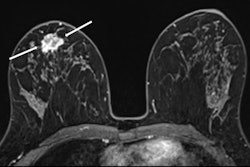
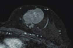
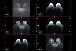
Final analysis included 80 women who underwent breast MRI after being diagnosed with architectural distortions on mammography between 2019 and 2022. Two independent radiologists recorded DCE-MRI lesion characteristics and IVIM parameters.
The researchers found no lesions on MRI in 33 women. For the remaining 47 women, 17 had benign lesions and 30 had malignant lesions. The combined approach achieved high performance marks in the study.
Performance of combined IVIM, DCE-MRI approach on characterizing architectural distortions | |
Measure | Score |
Sensitivity | 100% |
Specificity | 92% |
Negative predictive value | 100% |
Positive predictive value | 88.2% |
Accuracy | 95% |
The following DCE-MRI characteristics showed significant differences between benign and malignant architectural distortions for positive MRI findings: lesion diameter, shape, and margin of mass enhancement, distribution of nonmass enhancement, internal enhancement, and time signal intensity curve (p < 0.05). The researchers reported that internal enhancement and time signal intensity curve were independent risk predictors, with a combined area under the curve (AUC) of 0.969.
For IVIM parameters, pure diffusion coefficient, pseudo diffusion coefficient, and perfusion fraction showed significant differences between benign and malignant architectural distortions (all p < 0.05). This included AUC values of 0.908, 0.831, 0.680, and 0.761, respectively.
Finally, the team found that apparent diffusion coefficient and pseudo diffusion coefficient were independent risk predictors, with a combined AUC of 0.925. And combining DCE-MRI with IVIM achieved the highest AUC at 0.984.
The study authors highlighted that with this approach being reliable and non-invasive, it could help reduce unnecessary biopsies and surgeries.
The full study can be found here.