Dear Women's Imaging Insider,
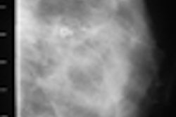
With its 3D images, digital breast tomosynthesis (DBT) can help mammographers see around overlapping breast tissue that might obscure pathology, making it a good adjunct to digital mammograms for women at high risk of breast cancer or with dense breast tissue.
But some have criticized the technology, citing its weakness in identifying microcalcifications, which digital mammograms tend to identify well.
Is DBT less effective than digital mammography in this regard? Researchers at Elizabeth Wende Breast Care conducted a study that compared the two modalities, and what they found may surprise you: Click here to read the article. As an Insider subscriber, you get access to the article before our other members.
Once you've read our feature, take a look at what else is going on in your Women's Imaging Digital Community:
- Find out why French researchers believe a second lumpectomy is a viable option for recurrent breast cancer.
- Read how the U.S Preventive Services Task Force's 2009 mammogram guidelines may put minorities and younger women at risk.
- Get the scoop on whether breast MRI makes surgical planning for patients with ductal carcinoma in situ more accurate.
- Learn what Canadian researchers have to say about the aggressiveness of interval breast cancers.
- Find out why radiation therapy has been tied to more-invasive recurrences after breast cancer surgery.
- Have your patients started asking for thyroid shields before mammograms? Learn why they might in this article.
As always, if you have a comment, report, or article idea to share about any aspect of women's imaging, I invite you to contact me.