Providing detailed instruction to maternal-fetal medicine physicians can improve the quality of fetal facial 3D ultrasound images, according to research published in the April edition of the Journal of Ultrasound in Medicine.
In a study that compared the performance of physicians who had received detailed instruction on 3D ultrasound volume manipulation to those who had instruction only on basic principles, researchers found that those with detailed instruction yielded significantly higher fetal profile and parallel-plane palate scores.
"Teaching physicians in a standardized way may help improve the use of [3D ultrasound] in clinical practice," wrote the team led by Dr. Gladys Ramos, of the University of California, San Diego.
While volume sonography has been explored for diagnosing a number of fetal abnormalities, clinical implementation has been challenging for a number of reasons, including training limitations, according to the researchers.
As a result, the study team sought to determine whether systematic instruction would help teach maternal-fetal medicine physicians with minimal 3D ultrasound experience how to display a diagnostic fetal profile and palate from 3D volumes (J Ultrasound Med, April 2011, Vol. 30:4, pp. 473-479).
The researchers recruited 10 physicians and randomly assigned them to two groups. Both groups were initially instructed on basic principles of 3D ultrasound volume manipulation using the 4D View program (GE Healthcare); the second group also received detailed, step-by-step 3D procedures on how to obtain the fetal profile.
Physicians in the first group (group A) were asked to display the fetal profile in five preselected fetal volumes, including one fetus with abnormalities. After receiving detailed instruction, group B physicians then also displayed the fetal profile in the same five volumes.
After the initial round of scanning, both groups were combined into one group and received the same detailed instruction provided previously to group B. Then the physicians were asked to review an additional five volumes.
In a later session, the researchers divided the physicians back into their respective groups and performed a similar exercise, this time for the display of the fetal palate in three-orthogonal-plane and parallel-plane images. Two experienced sonologists then reviewed all of the images in a blinded fashion for accuracy and clinical utility.
Group B turned in significantly higher fetal profile and parallel-plane palate scores than group A (p < 0.001).
Quality scoring for fetal profile
|
|||||||||||||||||||||||||||
Quality scoring for parallel-plane images of the fetal palate
|
|||||||||||||||||||||||||||
However, no significant difference was found between the groups for displaying the three-orthogonal-plane image of the palate or after additional training for either group, according to the researchers.
 |
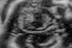
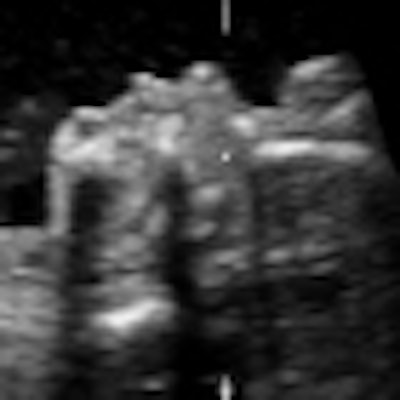
| Representative fetal profile images showing good-quality (above), acceptable-quality (below), and poor-quality (bottom) images. All images courtesy of the Journal of Ultrasound in Medicine. |
 |
 |
In other findings, the researchers did not detect a significant difference in mean display time between the groups. Evaluation time for abnormal profiles was longer than for normal profiles (p = 0.02).
"A systematic approach using clinical volumes provided maternal-fetal medicine physicians experience with practicing and training on personal computer workstations with a 'gamelike' atmosphere that stimulated the learner's interest and motivation for competition," the authors wrote. "An improved understanding of the importance of symmetry of the face was obtained from manipulating the volumes; this information is useful for both 2D and 3D evaluation of the face."
"We believe that additional practice (> 10 volumes) manipulating volumes is needed to consistently obtain adequate palate images using both three-orthogonal-plane and parallel-plane displays," they added.