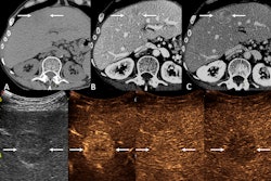
Contrast-enhanced ultrasound (CEUS) is a valuable tool for diagnosing hepatic nodules in patients with Fontan-associated liver disease, according to a study published May 26 in Radiology.
The finding is from a prospective study of the technique in 32 participants, with CEUS showing excellent interobserver agreement for nodule enhancement features and perfect agreement between two evaluators for nodule diagnoses, noted lead author David Reza Mittelstein, MD, PhD, of the University of California San Diego, and colleagues.
“A major strength of this study is that, to our knowledge, it is the first prospective cohort study to describe CEUS findings for hepatic nodules arising in [Fontan-associated liver disease],” the group wrote.
The Fontan procedure is an open-heart surgery performed in children to correct a heart defect. Since it was developed in the 1970s, the procedure has improved 20-year survival to 61% to 80%, yet over time it can cause multiorgan dysfunction due to elevated central venous pressure and decreased perfusion, the authors explained. In the liver, this manifests as a specific pattern of chronic congestive hepatopathy known as Fontan-associated liver disease (FALD).
FALD is characterized by progressive hepatic fibrosis, which may lead to cirrhosis and the formation of hepatic nodules, the researchers added. Patients with FALD are a small but unique population with characteristics that complicate CT or MRI, and thus the group aimed to evaluate CEUS as a potential alternative for diagnosing hepatic nodules.
The investigators enrolled 32 participants with FALD (mean age, 30 years old; 19 men) who underwent hepatic CEUS for nodule characterization between June 2018 and December 2023. Two CEUS-specialized physicians independently reviewed all studies, with interobserver variability quantified using Cohen κ. Biopsy diagnoses were obtained from pathologic examination for a subset of nodules.
 A visual abstract of the study.RSNA
A total of 35 hepatic nodules were identified and characterized. The two readers agreed on all 35 nodule diagnoses: 20 (57%) focal nodular hyperplasia-like lesions, seven (20%) regenerative nodules, five (14%) hemangiomas, and three (9%) possible malignancies. Interobserver agreement was 100% (κ = 1; no disagreements observed) for CEUS-guided nodule diagnoses. Interobserver agreement was also 100% for CEUS findings, except arterial phase hepatic enhancement pattern (mean κ = 0.92) and lesion echogenicity (mean κ = 0.55), according to the findings.
For five biopsied FALD nodules, CEUS diagnosis agreed with pathologic diagnosis, except for one diagnosis of possible malignancy that was biopsy-proven benign adenoma, the researchers added.
“In participants with FALD, there was excellent interobserver agreement in the evaluation of FALD nodules at hepatic CEUS, and CEUS diagnoses mostly agreed with pathologic diagnoses in the subset of cases with biopsies, including for malignant lesions and lesions requiring follow-up,” the group wrote.
In an accompanying editorial, Richard Barr, MD, PhD, of Northeastern Ohio Medical University in Rootstown, OH, wrote that the study, although small, confirms that CEUS enhancement features can help characterize nodules in FALD and that the enhancement features are similar to those of nodules associated with other chronic liver diseases.
“Additional multicenter studies that include cases of HCCs are needed to confirm these results,” Barr wrote.
The full study is available here.