Microwave ablation may be a suitable alternative to surgical resection when it comes to ultrasound-detected thyroid cancer, a study published April 2 in Radiology found.
Researchers led by Zhen-Long Zhao, MD, from China-Japan Friendship Hospital in Beijing found no significant difference in progression-free survival rates between patients receiving ablation or surgery. Additionally, microwave ablation led to less blood loss, shorter incision length, and shorter procedure and hospitalization durations.
“Microwave ablation is a feasible option for selected patients with multifocal T1N0M0 papillary thyroid carcinoma,” Zhao and colleagues wrote.
Microwave ablation has shown promise in previous studies for treating multifocal papillary thyroid carcinoma. The researchers highlighted that this method is minimally invasive and could spare healthy thyroid tissue. However, they also acknowledged that comparative studies are needed to measure microwave ablation’s efficacy against that of surgical resection.
The Zhao team compared both methods for preoperative ultrasound-detected T1N0M0 multifocal papillary thyroid carcinoma in terms of overall and one-, three-, and five-year progression-free survival rates and complication rates. It included retrospective data from 682 patients in 10 medical centers who received treatment between 2015 and 2021.
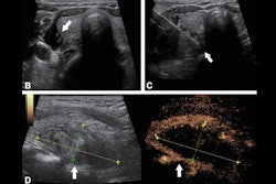
 Ultrasound images in a 57-year-old female patient with multifocal papillary thyroid carcinoma. (A) Routine ultrasound scan shows two hypoechoic papillary thyroid carcinoma nodules (arrows) in the left lobe. (B) Preablation contrast-enhanced ultrasound image shows hypoenhancement in the arterial phase (arrows). (C) Isolating fluid (black arrow) is used to protect the muscles beside the nodule (white arrow) during the ablation. (D) Postablation contrast-enhanced ultrasound image shows no enhancement in the original tumor zone (arrows). Image courtesy of the RSNA.
Of the total patients, 229 underwent microwave ablation and 453 underwent surgical resection and were observed for 20 months and 26 months, respectively.
Microwave ablation resulted in significantly less blood loss, shorter length of incision, and shorter procedure and hospitalization durations than surgical resection. Additionally, the team found no evidence of difference in progression-free survival rates in all time periods.
| Comparison between microwave ablation, surgical resection in thyroid cancer patients | |||
|---|---|---|---|
| |
Surgical resection |
Microwave ablation |
p-value |
| Estimated blood loss |
10 mL |
1 mL |
< 0.001 |
| Incision length |
7 cm |
0.4 cm |
< 0.001 |
| Procedure time |
90 |
30 |
< 0.001 |
| Hospitalization time |
5 days |
3 days |
< 0.001 |
| One-year survival rate |
98.5% |
98.7% |
0.39 |
| Three-year survival rate |
95.0% |
91.5% |
0.26 |
| Five-year survival rate |
83.1% |
77.2% |
0.36 |
The team also reported that permanent hoarseness (2.2%, p = 0.05) and hypoparathyroidism (4%, p = 0.005) were encountered only in the surgical resection group.
Finally, the researchers highlighted that microwave ablation was effective in managing the emergence of new tumors, observing no further tumor progression at the last follow-up.
“This outcome suggests that preserving normal thyroid tissue through microwave ablation does not notably heighten the risk of new tumors or negatively impact prognosis,” they added.
The study authors called for further validation of their results through prospective studies, writing that this would “solidify the role of microwave ablation in the treatment paradigm of multifocal papillary thyroid carcinoma.”
In an accompanying editorial, Christos Georgiades, MD, PhD, from Johns Hopkins University wrote that ablative modalities such as the one in the Zhao study should be considered due to the “indolent nature” of thyroid cancer.
“For patients in whom treatment is considered, there is an important next question that must be addressed: Which combination of clinical, morphologic, biochemical, and molecular data should favor treatment over surveillance and resection over ablation?” he added.
The full study can be found here.