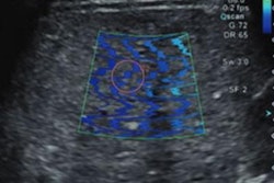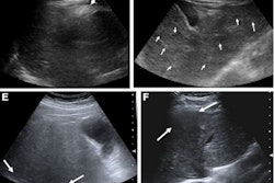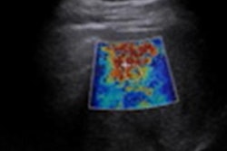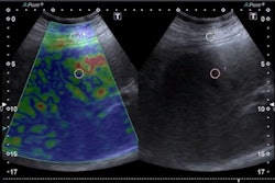
CHICAGO - COVID-19 patients who underwent ultrasound shear-wave elastography showed increased liver stiffness up to 10 months after infection, according to research presented December 1 at RSNA 2022.
 Dr. Firouzeh Heidari. Image courtesy of the RSNA.
The study adds to emerging evidence that COVID-19 infection may lead to liver injury that lasts well after the acute illness, said presenter Dr. Firouzeh Heidari of the Massachusetts General Hospital in Boston.
"We don't yet know if elevated liver stiffness observed after COVID-19 infection will lead to adverse patient outcomes," Heidari noted in an RSNA statement.
Liver stiffness is a marker of liver damage, such as fibrosis, which is the buildup of scar tissue in the liver. Over time, as healthy liver tissue diminishes, the liver can no longer function properly, and this can lead to liver failure.
Heidari and colleagues studied liver stiffness in 131 patients who underwent shear-wave elastography at Massachusetts General between 2019 and 2022. Patients were split into three groups based on when they underwent elastography and whether they tested positive for COVID-19.
One COVID-19-positive group included 31 patients who had a positive COVID-19 reverse transcription polymerase chain reaction (RT-PCR) test result at least 12 weeks before the elastography exam. Two control groups consisted of a random sample of 50 patients who underwent elastography during the COVID-19 pandemic and had a history of only negative COVID-19 PCR test results and a cohort of 50 patients who underwent an elastography exam prior to the COVID-19 pandemic.
In the COVID-positive group, elastography exams were performed an average of 44 weeks after a positive PCR test result.
After controlling for age, sex, and time period, a comparison of the elastography results among the groups showed that COVID-positive patients had a statistically significant higher liver stiffness than the control patients, and higher median liver stiffness (7.68 kPa) than pandemic control patients (5.99 kPa).
Additional research is warranted, Heidari said.
"We hope to enrich our existing database with additional patient data and a broader scope of co-variates to better understand the post-acute effects of COVID-19 within the liver," she stated.