Point-of-care lung ultrasound can rule out pulmonary congestion in critically ill patients with acute kidney injury, suggest findings published August 1 in the International Journal of Nursing Knowledge.
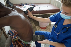
A team led by Bruna Barbeiro from the University of São Paulo in Brazil found that a single well-trained nurse achieved high specificity using lung ultrasound at the bedside in these patients, corresponding well to nursing reference terminology.
"Bedside nurse-performed [lung ultrasound] can strengthen clinical decision-making by quickly excluding pulmonary congestion, thereby informing volume-management interventions aligned with the NANDA-I taxonomy," the Barbeiro team wrote.
Previous studies from around the world have demonstrated success in training nonexperts, including nursing staff, to use ultrasound in settings that may not have easy access to medical care. Nurses use NANDA-I for reference to standardized nursing terminology.
The success of these training efforts with point-of-care ultrasound (POCUS) adds promise to the modality’s use in remote care settings.
Barbeiro and co-authors evaluated the diagnostic accuracy of lung ultrasound for finding pulmonary congestion in patients with acute kidney injury, with a nurse performing the exams. They also compared the ultrasound findings to defining characteristics from the NANDA-I nursing diagnosis of excess fluid volume.
For the study, a critical care nurse performed bedside lung ultrasound, which followed the bedside lung ultrasound in emergency protocol. Final analysis included a convenience sample of 64 intensive care unit patients with acute kidney injury in a general hospital in Brazil.
The researchers defined pulmonary congestion as ≥ 3 B-lines in ≥ 2 intercostal spaces per hemithorax. They also used radiological evidence of vascular congestion on chest radiograph or CT as the reference standard. The researchers found pulmonary congestion in 14 patients (21.9%).
The team reported the following findings:
Lung ultrasound sensitivity was 50% and specificity was 94%. Positive and negative predictive values were 70% and 87%, respectively.
Findings from lung ultrasound and radiological reference standards achieved substantial agreement (0.77).
The presence of ≥ 3 B-lines, a sonographic marker of interstitial fluid, corresponded to the characteristic of "pulmonary congestion" from NANDA-I diagnostic terminology.
Finally, the investigators reported no occurrences of adverse events from bedside lung ultrasound exams.
The results, including linking B-lines to defining characteristics of excess fluid volume, support POCUS’s use in nursing diagnostic reasoning, the study authors highlighted.
"Limited sensitivity and wide confidence intervals highlight the need for larger, multicenter studies with sufficient positive cases," they wrote.
The full study can be found here.