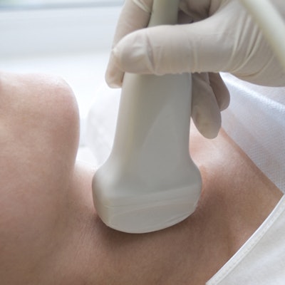
Transcutaneous laryngeal ultrasound shows potential as a noninvasive and accurate alternative to laryngoscopy for screening and postoperative review of vocal cord polyps, according to research published online June 23 in Ultrasound in Medicine & Biology.
In a study of 87 adult participants undergoing surgery at a hospital in China, transcutaneous laryngeal ultrasonography identified almost 90% of the polyps found on laryngoscopy. The modality was particularly effective in patients whose thyroid cartilage had little or no calcification.
"In this patient group, ultrasound can yield an accurate diagnosis, be noninvasive and safe, and be an effective supplement to laryngoscopy for the initial screening of vocal fold polyps and postoperative review," wrote the authors, led by Dr. Hua Wang from the department of ultrasound at the Second Affiliated Hospital of Xi'an Jiatong University in Xi'an.
As ultrasound becomes more prominent in clinical practice, studies have found the modality to be beneficial for visualizing numerous vocal cord issues, such as paralysis and nodules. However, few studies have evaluated the use of laryngeal ultrasound for vocal cord polyps, one of the most common benign vocal fold lesions seen in clinical practice, according to the authors.
The study included 87 participants undergoing surgery for vocal fold polyps at a single hospital in China. Before surgery, the patients underwent both laryngoscopy and transcutaneous laryngeal ultrasonography.
Ultrasound detected 81 polyps, compared with 92 detected by laryngoscopy. The rate of detection on ultrasound was higher for circular polyps (92%) than noncircular ones (71%), but the difference was not statistically significant.
The amount of thyroid cartilage had a substantial effect on ultrasound's ability to visualize the vocal folds of some patients. While the authors could visualize the folds for most patients, visibility decreased dramatically for patients with higher ratios of thyroid calcification to cartilage.
Notably, the authors visualized 100% of vocal folds for patients with little to no calcification and 90% of vocal folds in participants with a thyroid calcification-to-cartilage ratio of 34% to 66%. But the percentage of visualized folds fell to just 37.5% for patients with a calcification-to-cartilage ratio exceeding 66%.
"We found that thyroid cartilage calcification at the level of the glottis was the real influence on the vocal fold visualization," the authors wrote.
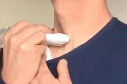
To help mitigate the effect of calcification and improve depth visualization, the authors placed the ultrasound probe at the side of the patients' neck at the level of the glottis. They also asked patients to hold their breath, which closed the vocal folds.
The authors intend to build upon their research in future studies with a larger number of participants and new imaging techniques. Their hope is to improve diagnostic accuracy even further, especially for noncircular polyps.
"We aim to find ways to improve the diagnostic accuracy of ultrasound for noncircular vocal fold polyps, develop ultrasound for the diagnosis of various benign and malignant lesions of vocal folds, and provide supportive data for voice and other related research," they wrote.