A risk-stratification system that includes scores from lung ultrasound scans may help emergency clinicians quickly make care decisions for patients with COVID-19, according to an article published on May 9 in Emergency Medicine Australasia.
The protocol, called cardiopulmonary limited ultrasound examination (CLUE), is an established method to quickly get data about a patient's heart and lung health. The authors slightly altered the emergency medicine tool to reflect the needs of patients with the novel coronavirus.
"CLUE protocol involves an anatomical parameter, lung ultrasound scoring system, and a physiological parameter, oxygen at the time of examination, to aid emergency clinicians in making disposition decisions," wrote the authors, led by Dr. Vijay Manivel from the department of emergency medicine at Nepean Hospital in Kingswood, Australia.
Lung ultrasound scans have repeatedly proved helpful for clinicians needing to make admission, discharge, and treatment decisions for patients with COVID-19, but so far, there's been no standardized risk stratification system. The proposed CLUE protocol seeks to change that for physicians in Australia and New Zealand who are currently preparing to see a wave of patients with COVID-19.
"While Australia and New Zealand prepare for a figurative tsunami of highly infectious patients, we anticipate that a protocolized use of bedside [lung ultrasound scans] by emergency clinicians in COVID-19 patients could alleviate some of the radiological resource burden expected," the authors wrote.
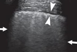
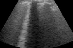
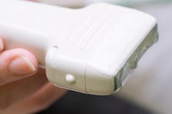
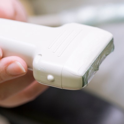
The CLUE protocol includes a 12-zone lung ultrasound scan. During the scan, a trained emergency physician waves a transducer across a patient's anterior chest wall, lateral chest wall, and posterior chest wall, looking for hallmark signs of COVID-19.
For each zone, the clinician would calculate a number using the lung ultrasound scoring system (LUSS):
- LUSS 0 -- A-lines or one to two B-lines, a smooth, thin pleural line
- LUSS 1 -- More than two, discrete B-lines, an irregular or thickened pleura
- LUSS 2 -- Confluent B-lines, subpleural consolidations less than 1 cm in height
- LUSS 3 -- Consolidations more than 1 cm in height, air bronchogram, vascularity
The CLUE protocol then takes the cumulative LUSS score for each of the 12 zones and adds it to other patient factors to classify the severity of the patient and the suggested treatment.
| CLUE protocol for patients with COVID-19 | |||
| Risk classification | LUSS Score | Supplemental oxygen | Suggested treatment(s) |
| Normal | 0 | No | • Send patient home |
| Yes | • Consider alternate cause | ||
| Mild | 1-5 | No | • Send patient home with follow-up monitoring |
| Yes | • Send patient home with pulse oximetry monitoring and/or oxygen
• Consider ward admission • Involve respiratory/ICU physician |
||
| Moderate | 6-15 | No | • Send patient home with pulse oximetry monitoring and/or oxygen
• Consider ward admission • Involve respiratory/ICU physician |
| Yes | • Admit to ward
• Consider ICU admission • Involve respiratory/ICU physician |
||
| Severe | 16+ | No | • Admit to ward
• Involve respiratory/ICU physician |
| Yes | • Admit to ICU
• Involve respiratory/ICU physician |
||
The authors cautioned that the CLUE protocol is only a suggestion and that clinicians should also take into account other patient factors, such as heart disease, when making patient diagnostic and triage decisions. They are currently conducting a multicenter trial to test the system in emergency departments throughout Australia and New Zealand.
The researchers hope that the CLUE protocol unifies patient care and reduces the need for supplemental radiology procedures.
"This protocol will aid the clinician to make rapid and appropriate bedside clinical decisions, potentially decrease reliance on chest x-rays or CT scans, and aid disposition planning from the emergency department," they concluded.