More than a dozen case reports and studies from around the world showed consistent lung ultrasound scan findings in patients with COVID-19, according to a review published on May 25 in Ultrasound in Medicine and Biology.
The review, conducted by two radiologists from the ultrasound research lab at the University of Pennsylvania, demonstrated the feasibility of lung ultrasound as a first-line resource for patients with the novel coronavirus. The research is part of the lab's ongoing effort to standardize ultrasound features and quantify and automate ultrasound findings.
"Lung ultrasound has emerged as a strong first-line imaging modality in emergency settings for diagnosing COVID-19 as well as tracking disease progression," study co-author Dr. Laith Sultan told AuntMinnie.com. "The findings from all the studies we reviewed from around the globe showed high sensitivity comparable to CT-scan."
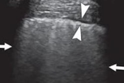
Sultan and colleague Chandra Sehgal, PhD, reviewed lung ultrasound findings of adult and pediatric patients with COVID-19 from 14 sources, including case reports and prior studies. They found common features of COVID-19 on ultrasound scans, which included multiple B-line patterns with spared areas and thickened, irregular pleural lines.
Many of the ultrasound findings were bilateral and posterobasal predominant, they noted. Scans also revealed subpleural consolidations, which can be associated with discrete, localized pleural effusion.
The hallmark signs of COVID-19 were consistent in Europe and Asia, as well as present in emerging research from Canada and Turkey. The findings also appeared in children and pregnant patients.
"One of the things that really impressed me is how characteristic patterns of lung ultrasound findings for COVID-19 patients surfaced from around the globe in such a short duration," Sultan said. "Particularly, I was impressed with the Italian experience and how organized and proactive were the medical teams over there."
More specific findings related to COVID-19 included that patients with severe, progressive disease often had the presence of alveolar consolidation with a tissue-like appearance, as well as dynamic and static air bronchograms on ultrasound scans.
In addition, a research team from China used color Doppler flow imaging ultrasound scans to look at blood flow in the lungs of patients. The team found signs of lack of blood flow signals in subpleural consolidations, which could have resulted from larger, systemic COVID-19 effects.
"Looking at the findings, some features really surprised me," Sultan said. "The absence of Doppler signal, which is opposite to what one can expect in inflammatory lung changes, is really a unique finding for COVID-19."
While the results are promising, the researchers emphasized the need for a standardized lexicon for ultrasound features related to COVID-19, similar to the language used for sonography for breast, liver, and thyroid cancers. They're also working with teams in Italy and Spain on machine learning that may one day be able to help physicians diagnose COVID-19.
"Penn's Ultrasound research lab started working on multiple projects that focus on the automation of sonographic findings to reduce user training and user variability," Sultan said. "These projects involve the use of computer image analysis and machine learning methods."

![Examples of ultrasound findings and techniques. (A) Images in a 39-year-old male patient with a mass in the left thigh. The mass is heterogeneous on the B-mode US image (compared with the patient in D) and showed increased microvascularity (superb microvascular imaging [SMI]) and shear-wave elastography (SWE) values. Undifferentiated pleomorphic sarcoma was diagnosed at biopsy (with pleomorphic rhabdomyosarcoma in surgical specimen). (B) Images in an 18-year-old male patient with a mass in the left leg. The mass is hypoechoic on the B-mode image, with no other findings suggestive of malignancy. The lesion is in contact with the cortex of the tibia, which is slightly irregular. CT revealed a doubtful anteromedial tibial erosion. The microvascular study demonstrated high vascularization, suggestive of malignancy. Periosteal Ewing sarcoma was diagnosed with both histologic and immunohistochemical confirmation. (C) Images in a 69-year-old female patient with a lump growing on the outside of the left leg. Multiple SWE examinations were performed (please note the high values obtained in the measurements, whereas the color map highlights the stiffness relative to adjacent tissues). SMI showed areas of increased vascularization to target for sampling. Undifferentiated spindle cell sarcoma was diagnosed at biopsy, with residual leiomyosarcoma in the surgical specimen after neoadjuvant therapy. (D) Images in a 56-year-old female patient with a mass in the right thigh. The mass is heterogeneous at both B-mode ultrasound (similar to patient A) and MRI (coronal T2-weighted spectral attenuated inversion recovery [SPAIR]; T1-weighted pre-contrast and postcontrast imaging), which even shows uptake after the administration of paramagnetic contrast material, which is traditionally suggestive of malignancy. Low values at SMI and elastography are suggestive of benignity. Spindle cell lipoma was diagnosed at biopsy, with atypical spindle cell lipomatous tumor in the surgical specimen.](https://img.auntminnie.com/mindful/smg/workspaces/default/uploads/2026/08/images-radiol250278fig2.APCFLSvX6p.jpg?auto=format%2Ccompress&fit=crop&h=100&q=70&w=100)


![Examples of ultrasound findings and techniques. (A) Images in a 39-year-old male patient with a mass in the left thigh. The mass is heterogeneous on the B-mode US image (compared with the patient in D) and showed increased microvascularity (superb microvascular imaging [SMI]) and shear-wave elastography (SWE) values. Undifferentiated pleomorphic sarcoma was diagnosed at biopsy (with pleomorphic rhabdomyosarcoma in surgical specimen). (B) Images in an 18-year-old male patient with a mass in the left leg. The mass is hypoechoic on the B-mode image, with no other findings suggestive of malignancy. The lesion is in contact with the cortex of the tibia, which is slightly irregular. CT revealed a doubtful anteromedial tibial erosion. The microvascular study demonstrated high vascularization, suggestive of malignancy. Periosteal Ewing sarcoma was diagnosed with both histologic and immunohistochemical confirmation. (C) Images in a 69-year-old female patient with a lump growing on the outside of the left leg. Multiple SWE examinations were performed (please note the high values obtained in the measurements, whereas the color map highlights the stiffness relative to adjacent tissues). SMI showed areas of increased vascularization to target for sampling. Undifferentiated spindle cell sarcoma was diagnosed at biopsy, with residual leiomyosarcoma in the surgical specimen after neoadjuvant therapy. (D) Images in a 56-year-old female patient with a mass in the right thigh. The mass is heterogeneous at both B-mode ultrasound (similar to patient A) and MRI (coronal T2-weighted spectral attenuated inversion recovery [SPAIR]; T1-weighted pre-contrast and postcontrast imaging), which even shows uptake after the administration of paramagnetic contrast material, which is traditionally suggestive of malignancy. Low values at SMI and elastography are suggestive of benignity. Spindle cell lipoma was diagnosed at biopsy, with atypical spindle cell lipomatous tumor in the surgical specimen.](https://img.auntminnie.com/mindful/smg/workspaces/default/uploads/2026/08/images-radiol250278fig2.APCFLSvX6p.jpg?auto=format%2Ccompress&fit=crop&h=112&q=70&w=112)