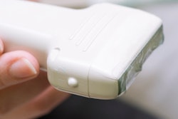
Ultrasound is an effective point-of-care imaging modality for diagnosing necrotizing fasciitis, a condition that can result in death in almost 20% of patients if not caught early, according to a study published online April 26 in Ultrasound in Medicine and Biology.
The modality can identify fluid accumulation in the fascia, giving clinicians the information they need to better diagnose and treat the condition, wrote a team led by Dr. Chun-Nan Lin of Chang Gung Memorial Hospital in Chiayi, Taiwan.
"The improved prediction of prognosis might help physicians to explain the severity of the disease and accelerate early surgical intervention," the authors wrote.
Known popularly as "flesh-eating disease," necrotizing fasciitis is a serious soft-tissue infection with a mortality rate of almost 20%. Its clinical presentation is fever, pain that's out of proportion with skin findings, a popping or crackling sound in the joint, fluid-filled sacs or lesions, necrosis, or bruising.
Early surgical intervention can reduce the mortality rate of necrotizing fasciitis, so early diagnosis is crucial, the team wrote. The condition is typically assessed using CT or MRI, but these modalities may not be immediately available when a patient presents in the emergency department.
That's where ultrasound comes in, according to the researchers.
"Ultrasonography is a convenient, cost-effective, and noninvasive tool, and its utility may enable prompt diagnosis of necrotizing fasciitis," they wrote.
Ultrasound findings for the disease include irregularity or thickening of the deep fascia, abnormal fluid collection along the deep fascial plane, and trapped air under the skin. Lin's team investigated the relationship between the ultrasound finding of fluid accumulation, and the diagnosis and prognosis of necrotizing fasciitis.
The study included 95 patients. Of these, 48 had necrotizing fasciitis and 47 did not. Four of the 48 patients with necrotizing fasciitis died due to disease progression and despite surgical intervention.
Lin and colleagues found that 85.4% of patients with necrotizing fasciitis had fluid accumulation, compared with 55.3% of those patients without the disease (p = 0.001). The best cutoff point of fluid accumulation to diagnose necrotizing fasciitis in terms of accuracy was 2 mm.
| Ultrasound performance for diagnosing necrotizing fasciitis by depth of fluid accumulation | ||
| Measure | 1 mm | 2 mm |
| Accuracy | 68.7% | 72.7% |
| Sensitivity | 86.5% | 75% |
| Specificity | 48.9% | 70.2% |
| Positive predictive value | 65.2% | 71.7% |
| Negative predictive value | 76.7% | 72.7% |
The group also found that patients with necrotizing fasciitis had longer hospital stays compared with those who did not have the disease (39 versus 23 days).
"Our study showed that ultrasonographic finding of fluid accumulation along the deep fascia may facilitate the diagnosis and prognosis of necrotizing fasciitis," Lin's team concluded. "In regard to the prognosis of necrotizing fasciitis, when fluid accumulation was present along the deep fascia, patients with [the disease] had longer lengths of hospital stays and were at risk of amputation or mortality. Therefore, ultrasonography can be used as a point-of-care imaging tool to help clinicians make critical decisions on the diagnosis and prognosis of necrotizing fasciitis."