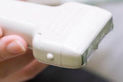
Point-of-care ultrasound (POCUS) shows promise as a tool for emergency responders to help predict survival and guide interventions for cardiac arrests experienced outside of the hospital, according to research presented at the recent American Institute of Ultrasound in Medicine (AIUM) meeting in New York City.
POCUS is becoming more commonplace as a tool used for resuscitation of patients in cardiac arrest in the hospital, but there are not much data on its use outside of the hospital setting, presenter Dr. Katherine German of the University of Pittsburgh told session attendees.
"We know that out-of-hospital cardiac arrest survival is low, and we were curious whether point-of-care ultrasound could be a useful tool to predict survival and guide therapy or the decision to transport in the field," German said.
A helpful tool
German and colleagues explored the feasibility of POCUS when used by emergency staff in patients experiencing cardiac arrest outside of hospitals. A portable ultrasound unit (iViz, Fujifilm SonoSite) was used in vehicles as part of the Pittsburgh Emergency Medical Services (EMS) system; all participating physicians received POCUS education and watched an instructional video on how to use the device. Physicians provided radio and in-person consultation to Pittsburgh city paramedics.
The field staff used ultrasound to identify cardiac motion during pulse checks -- without interrupting resuscitation -- and to guide management of the situation at the physicians' discretion. The POCUS users recorded data from still images and six-second video clips, as well as through electronic charting within the patient care report using emsCharts, a tool for air and ground emergency medical personnel.
The study's primary purpose was to track the frequency of POCUS use during out-of-hospital cardiac arrest events; the secondary purpose was to have an expert faculty reviewer analyze images acquired during these events and to identify any barriers to the use of the technology.
German's group included 348 field responses that occurred between November 2016 and March 2017. Of these, 127 were out-of-hospital cardiac arrest. Field doctors used POCUS in 56 (44%) of the out-of-hospital cardiac arrest cases, with 48 (85%) still images and 34 (61%) video images recorded and reviewed.
Agreement between interpretations of emergency responder physicians and those of expert reviewers was found in 91% of video cases. Responders noted the following reasons for not using POCUS:
- Return of spontaneous circulation (ROSC) close to physician arrival (30%)
- Emergency personnel busy with other interventions (21%)
- Provider preference (23%)
- Mechanical issues (6%)
- Cessation of resuscitation per advanced directives (6%)
In the field
German did note some of the study's limitations, including the user dependency of ultrasound, as well as the fact that POCUS use was optional and that interpretations were recorded after the technology was used in the field. She also cited the small sample size of video images.
However, overall, POCUS showed promise as a tool for dealing with cardiac arrest in the field, she concluded.
"POCUS use by novice physician ultrasonographers to detect cardiac wall motion in out-of-hospital cardiac arrest is not only feasible, it also correlates with expert interpretation," German said. "Barriers to its use can be addressed through educational interventions and increased familiarity with the device."


![Examples of ultrasound findings and techniques. (A) Images in a 39-year-old male patient with a mass in the left thigh. The mass is heterogeneous on the B-mode US image (compared with the patient in D) and showed increased microvascularity (superb microvascular imaging [SMI]) and shear-wave elastography (SWE) values. Undifferentiated pleomorphic sarcoma was diagnosed at biopsy (with pleomorphic rhabdomyosarcoma in surgical specimen). (B) Images in an 18-year-old male patient with a mass in the left leg. The mass is hypoechoic on the B-mode image, with no other findings suggestive of malignancy. The lesion is in contact with the cortex of the tibia, which is slightly irregular. CT revealed a doubtful anteromedial tibial erosion. The microvascular study demonstrated high vascularization, suggestive of malignancy. Periosteal Ewing sarcoma was diagnosed with both histologic and immunohistochemical confirmation. (C) Images in a 69-year-old female patient with a lump growing on the outside of the left leg. Multiple SWE examinations were performed (please note the high values obtained in the measurements, whereas the color map highlights the stiffness relative to adjacent tissues). SMI showed areas of increased vascularization to target for sampling. Undifferentiated spindle cell sarcoma was diagnosed at biopsy, with residual leiomyosarcoma in the surgical specimen after neoadjuvant therapy. (D) Images in a 56-year-old female patient with a mass in the right thigh. The mass is heterogeneous at both B-mode ultrasound (similar to patient A) and MRI (coronal T2-weighted spectral attenuated inversion recovery [SPAIR]; T1-weighted pre-contrast and postcontrast imaging), which even shows uptake after the administration of paramagnetic contrast material, which is traditionally suggestive of malignancy. Low values at SMI and elastography are suggestive of benignity. Spindle cell lipoma was diagnosed at biopsy, with atypical spindle cell lipomatous tumor in the surgical specimen.](https://img.auntminnie.com/mindful/smg/workspaces/default/uploads/2026/08/images-radiol250278fig2.APCFLSvX6p.jpg?auto=format%2Ccompress&fit=crop&h=100&q=70&w=100)


![Examples of ultrasound findings and techniques. (A) Images in a 39-year-old male patient with a mass in the left thigh. The mass is heterogeneous on the B-mode US image (compared with the patient in D) and showed increased microvascularity (superb microvascular imaging [SMI]) and shear-wave elastography (SWE) values. Undifferentiated pleomorphic sarcoma was diagnosed at biopsy (with pleomorphic rhabdomyosarcoma in surgical specimen). (B) Images in an 18-year-old male patient with a mass in the left leg. The mass is hypoechoic on the B-mode image, with no other findings suggestive of malignancy. The lesion is in contact with the cortex of the tibia, which is slightly irregular. CT revealed a doubtful anteromedial tibial erosion. The microvascular study demonstrated high vascularization, suggestive of malignancy. Periosteal Ewing sarcoma was diagnosed with both histologic and immunohistochemical confirmation. (C) Images in a 69-year-old female patient with a lump growing on the outside of the left leg. Multiple SWE examinations were performed (please note the high values obtained in the measurements, whereas the color map highlights the stiffness relative to adjacent tissues). SMI showed areas of increased vascularization to target for sampling. Undifferentiated spindle cell sarcoma was diagnosed at biopsy, with residual leiomyosarcoma in the surgical specimen after neoadjuvant therapy. (D) Images in a 56-year-old female patient with a mass in the right thigh. The mass is heterogeneous at both B-mode ultrasound (similar to patient A) and MRI (coronal T2-weighted spectral attenuated inversion recovery [SPAIR]; T1-weighted pre-contrast and postcontrast imaging), which even shows uptake after the administration of paramagnetic contrast material, which is traditionally suggestive of malignancy. Low values at SMI and elastography are suggestive of benignity. Spindle cell lipoma was diagnosed at biopsy, with atypical spindle cell lipomatous tumor in the surgical specimen.](https://img.auntminnie.com/mindful/smg/workspaces/default/uploads/2026/08/images-radiol250278fig2.APCFLSvX6p.jpg?auto=format%2Ccompress&fit=crop&h=112&q=70&w=112)