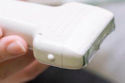
Do you want to help residents brush up on their point-of-care (POCUS) ultrasound skills? Try turning educational content into a competitive game, according to a study published in the November issue of the Journal of Ultrasound in Medicine.
Generation Y students -- also known as millennials -- reportedly learn better in groups using hands-on activities and new technology, so using gamification techniques to teach medical skills may be particularly effective with residents and medical students, wrote a team led by Dr. Andrew Liteplo of Massachusetts General Hospital in Boston.
Since 2011, the Society for Academic Emergency Medicine has held at its annual meeting a competitive games-based event called SonoGames that's intended to educate emergency medicine residents about point-of-care ultrasound.
The event has been quite successful, according Liteplo and colleagues.
"SonoGames [has] positively affected resident perceptions and understanding of ultrasound across clinical practice, knowledge and competence, and enthusiasm," the group wrote.
The SonoGames event consists of three rounds of play that assess hand-eye coordination, motor task mastery, spatial recognition, interpretation of images, and understanding of anatomy skills for point-of-care ultrasound. Round 1 consists of multiple choice questions in regard to ultrasound clips, while rounds 2 and 3 feature hands-on scanning tests, the researchers wrote (J Ultrasound Med, November 2018, Vol. 37:11, pp. 2491-2496).
Liteplo and colleagues investigated the effect of the May 2016 event on the immediate learning and long-term education of those who participated. The researchers created two surveys, one for emergency medicine residents and another for program directors, which they sent out via email in June 2016. Seventy-three residents and 42 directors participated.
The surveys used a five-point scale to elicit overall perceptions and attitudes about ultrasound, descriptions of any changes in knowledge and competency after the games, effects of the games on clinical use, and impact of the game format on education.
Of all the participants, 94% stated that SonoGames was effective as an educational tool. The researchers also found the following:
- 87% of survey respondents said their enthusiasm for ultrasound increased.
- 81% reported that SonoGames increased their ultrasound knowledge.
- 75% said that SonoGames taught them material they then incorporated into their clinical practice.
- 61% said their clinical use of ultrasound increased.
- 42% said that participating in SonoGames inspired them to pursue an ultrasound-based career.
Survey participants who moved further through the SonoGames rounds had more positive responses, Liteplo and colleagues noted.
"[Participants] who advanced to round 2 found the event to be more effective at education, motivation, and enthusiasm than those who left after round 1," the group wrote. "This finding was not surprising, as hands-on games are more interactive and fun than multiple choice questions."
The study results suggest that events like SonoGames could "serve as a model for resident teaching and learning across other modalities," Liteplo and colleagues wrote.
"This study supports the use of gamification as an innovative, impactful tool that has applicability in training learners of all levels on a national scale," they concluded.