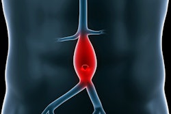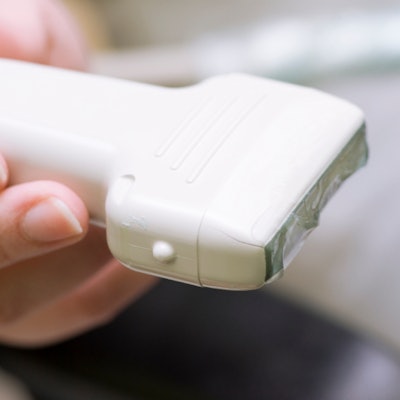
With a bit of training, medical students using point-of-care ultrasound (POCUS) can identify more abdominal aortic aneurysms (AAAs) than vascular surgeons can by screening for the condition via physical exam, according to a study published online May 17 in the Annals of Vascular Surgery.
The findings not only confirm ultrasound's efficacy for this application but also suggest a new resource for AAA screening in an era when medical staff can be stretched thin, wrote the team led by Trinh Mai from the University of Ottawa.
"Screening for AAA has been found to be largely underutilized in the U.S., with only 15% of eligible and at-risk individuals receiving screening examinations," the researchers wrote. "To mitigate obstacles associated with access to screening [such as staff and device availability] ... the introduction of point-of-care ultrasound training at the medical student level, and its wide-scale implementation as an extension to physical examination, may lead to improved detection of AAA."
Ultrasound training
When untreated, abdominal aortic aneurysms have a mortality rate as high as 85% to 90%, Mai and colleagues wrote. Physical examination has low sensitivity and specificity, even when experienced physicians perform it.
Ultrasound is much more effective and is currently the primary imaging tool used to screen for the condition, but its widespread use for this application has been hindered by cost and the availability of appropriately trained technicians.
However, there's mounting evidence that medical personnel with no previous training can effectively use ultrasound to screen for AAA, especially with point-of-care ultrasound.
"The objective of our study was to determine whether a medical student, after a short training period, could reliably screen for abdominal aortic aneurysms," the group wrote. "We compared the test characteristics of point-of-care ultrasound performed by a medical student versus physical examination by vascular surgeons and a gold standard reference scan."
For the study, the researchers included data from 57 patients who presented to an outpatient vascular surgery clinic between October 2015 and March 2016 to be screened for abdominal aortic aneurysm. A vascular surgeon evaluated each patient via physical exam, followed by a second-year medical student using POCUS; patients then underwent a reference CT or vascular sonographer-performed ultrasound exam within three months of this initial screening visit.
The student had three hours of training with POCUS for detecting and measuring AAA. The training included a theoretical component led by a vascular sonographer and a practical component taught by an emergency physician, during which the student practiced ultrasound scanning under direct guidance.
After this initial training, the student practiced using ultrasound on the abdominal aortas of 60 patients with and without the condition. The student then completed a competency assessment before beginning to scan study participants, Mai and colleagues wrote.
Most of the patients (61%) were men, and the median patient age was 71. Reference exams identified 16 abdominal aortic aneurysms with a mean maximum aortic diameter of 29.5 mm. Of these, the vascular surgeon identified 11, with two false positives, for a sensitivity rate of 66.7% and a specificity rate of 94.4%. The average time to conduct the physical exam was 35 seconds.
Medical students using point-of-care ultrasound found 15 of the 16 abdominal aortic aneurysms with a mean maximum aortic diameter of 28 mm, for a sensitivity rate of 93.3% and a specificity rate of 100%. The average time to conduct the ultrasound exam was four minutes, the group wrote.
| Physical exam vs. POCUS for AAA screening | ||
| Measure | Physical exam | Point-of-care ultrasound |
| Sensitivity | 68.8% | 93.8% |
| Specificity | 93.9% | 100% |
| Positive predictive value | 81.8% | 100% |
"The results of our study, as well as several others, have proven that physical examination is relatively unreliable for the detection of abdominal aortic aneurysms," the researchers noted. "Screening for abdominal aortic aneurysms may be accurately and efficiently performed by a novice operator using point-of-care ultrasound with relatively limited training and experience."
Core curriculum?
Because point-of-care ultrasound appears to be an effective tool for identifying AAA, perhaps its use should be more formally incorporated into the medical school curriculum, Mai's team suggested.
"This study raises the question of whether we should be changing the way we are teaching medical students," the group wrote. "With the increased prevalence and use of ultrasound in medical practice, perhaps we should consider teaching focused ultrasound for abdominal aortic aneurysm detection as a standard in the medical school curriculum."