Researchers from the University of Arkansas in Fayetteville have published results from a study testing a new ultrasound imaging method for the detection and diagnosis of congenital heart disease in infants and children.
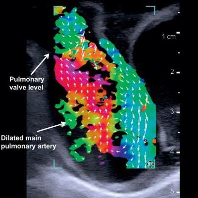
The new technology, called vector flow, creates images of the internal structure and blood flow of children's hearts. It was used for the first time in humans at the Arkansas Children's Hospital in Little Rock, according to a team led by Dr. Morten Jensen, PhD. The study was published March 5 in Progress in Pediatric Cardiology.
About 1% of babies are born with congenital heart defects. Pediatric cardiologists identify congenital heart disease using echocardiography and other processes based on ultrasound, the researchers wrote. Although effective, ultrasound can't accurately obtain details about blood flow within the heart.
The team used an ultrasound scanner with vector flow imaging to image the hearts of two three-month-old babies, one with a healthy heart and one with congenital heart disease. The technology allowed for complete transthoracic imaging of tissue and blood flow at a depth of 6.5 cm; abnormal flow and cardiac anomalies were clearly visualized in the child with congenital heart disease.
Vector flow imaging demonstrates swirl of blood flow within the dilated main pulmonary artery of a pig. Image courtesy of Dr. Morten Jensen, PhD."Vector flow imaging technology is not yet possible in adults, but we have demonstrated that it is feasible in pediatric patients," Jensen said in a statement released by the university April 3. "Our group demonstrated that this commercially available technology can be used as a bedside imaging method, providing advanced detail of blood flow patterns within cardiac chambers, across valves, and in the great arteries."







![Examples of ultrasound findings and techniques. (A) Images in a 39-year-old male patient with a mass in the left thigh. The mass is heterogeneous on the B-mode US image (compared with the patient in D) and showed increased microvascularity (superb microvascular imaging [SMI]) and shear-wave elastography (SWE) values. Undifferentiated pleomorphic sarcoma was diagnosed at biopsy (with pleomorphic rhabdomyosarcoma in surgical specimen). (B) Images in an 18-year-old male patient with a mass in the left leg. The mass is hypoechoic on the B-mode image, with no other findings suggestive of malignancy. The lesion is in contact with the cortex of the tibia, which is slightly irregular. CT revealed a doubtful anteromedial tibial erosion. The microvascular study demonstrated high vascularization, suggestive of malignancy. Periosteal Ewing sarcoma was diagnosed with both histologic and immunohistochemical confirmation. (C) Images in a 69-year-old female patient with a lump growing on the outside of the left leg. Multiple SWE examinations were performed (please note the high values obtained in the measurements, whereas the color map highlights the stiffness relative to adjacent tissues). SMI showed areas of increased vascularization to target for sampling. Undifferentiated spindle cell sarcoma was diagnosed at biopsy, with residual leiomyosarcoma in the surgical specimen after neoadjuvant therapy. (D) Images in a 56-year-old female patient with a mass in the right thigh. The mass is heterogeneous at both B-mode ultrasound (similar to patient A) and MRI (coronal T2-weighted spectral attenuated inversion recovery [SPAIR]; T1-weighted pre-contrast and postcontrast imaging), which even shows uptake after the administration of paramagnetic contrast material, which is traditionally suggestive of malignancy. Low values at SMI and elastography are suggestive of benignity. Spindle cell lipoma was diagnosed at biopsy, with atypical spindle cell lipomatous tumor in the surgical specimen.](https://img.auntminnie.com/mindful/smg/workspaces/default/uploads/2026/08/images-radiol250278fig2.APCFLSvX6p.jpg?auto=format%2Ccompress&fit=crop&h=112&q=70&w=112)