(Radiology Review) Obstetricians at the Weill Medical College of Cornell University in New York evaluated the significance of umbilical cord thickness in aneuploid fetuses, and the results were published in the September issue of the Journal of Ultrasound in Medicine (September 2004, Vol. 23, pp. 1177-1183).
The early detection of fetal aneuploidy remains an important challenge for perinatologists and is important for improved patient management, according to Dr. Mladen Predanic and colleagues.
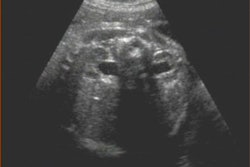
Nuchal translucency screening was performed at the hospital between 10 and 14 weeks' gestational age, while fetal morphology was evaluated between 18 and 23 weeks' gestational age. A retrospective cross-sectional study was performed of 56 fetuses and neonates with abnormal karyotypes diagnosed in utero or at birth during a two-year period. Forty-six fetuses had numerical chromosomal abnormalities. There were 26 fetuses with adequate sonographic imaging of the umbilical cord, and the measured thickness was compared with previously established umbilical cord thickness nomograms.
For fetuses 18 weeks' gestation or more, measurement of umbilical cord diameter was performed at a level less than 0.5 cm from the abdominal wall insertion. When the gestational age was between 14 and 18 weeks' gestation, a transverse section of cord was measured at the level of the wall insertion or at the level of the cord-free loop.
"Measurement calipers were placed at the outer edges of the umbilical cord to include umbilical cord vessels and the surrounding Wharton jelly," the authors wrote. Also, the number of umbilical cord vessels and amount of Wharton jelly was assessed. Ultrasound imaging was performed using a 6-MHz transabdominal transducer that had multiple frequency choice and harmonic capability for improved image quality.
Results demonstrated a cord thickness greater than the 95th percentile in 53.8% of fetuses: 57.8% of fetuses with trisomy 21, and 50% of subjects with trisomy 18. "The largest number of aneuploid fetuses with thick umbilical cords (87.5%) was observed between 16 and 17 gestational weeks," they wrote. Sixty-six percent of fetuses had abnormal genetic testing in the first trimester.
The authors suggested that the mechanisms that caused NT edema might also be responsible for umbilical cord swelling in aneuploid fetuses. Because the number of thick umbilical cords decreased with gestational age, they speculated that the increased amount of Wharton jelly associated with abnormal karyotypes resolved as pregnancies progressed. Just as NT edema is a transient abnormal finding, umbilical cord thickening might also be age-dependent. They found only one case of trisomy 18 with a cord thickness greater than the 95th percentile after 19 weeks' gestational age.
The authors concluded that aneuploid fetuses had thicker umbilical cords than euploid fetuses, and that the percentage of fetuses with thick umbilical cords was greater in this study than had been reported previously.
"Fetal Aneuploidy and Umbilical Cord Thickness Measured Between 14 and 23 Weeks' Gestational Age"
Predanic, M., et al
Division of Maternal-Fetal Medicine
Department of Obstetrics and Gynecology
Weill Medical College of Cornell University
525 E 68th Street, Suite M-704, New York, NY 10021 USA
J Ultrasound Med 2004 (September); 23: 1177-1183
By Radiology Review
October 21, 2004
Copyright © 2004 AuntMinnie.com