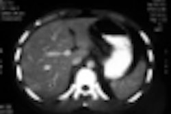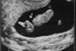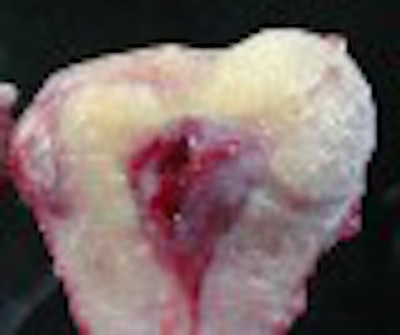
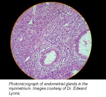
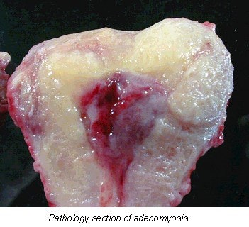
Many women who present for ultrasound examination with abnormal premenopausal vaginal bleeding are being misdiagnosed as having multiple small fibroids, according to a Canadian professor. In actuality, these patients may have adenomyosis, a benign disease of the uterus in which endometrial glands and stroma are found within the myometrium.
Dr. Edward Lyons, professor of radiology, obstetrics, and gynecology at the University of Manitoba in Winnipeg, Canada, presented the results of his research at the 2001 Australian Society for Ultrasound in Medicine meeting in Sydney.
"The most common gynecological causes of abnormal premenopausal bleeding are adenomyosis, submucosal fibroids, polyps, dysfunctional bleeding, and, less likely, endometrial carcinoma," he said.
Until recently, an accurate diagnosis of adenomyosis could be made only by microscopic examination of uterine specimens obtained during surgery, or, less often, during biopsy. The exact prevalence of the disease is not clear. Some studies estimate that 20% of women have adenomyosis; however, with careful microscopic analysis of multiple myometrial samples from an individual uterine specimen, the prevalence increases to as high as 65%.
 |
Lyons said his department was unfamiliar with the ultrasound appearances of adenomyosis, but a 1999 investigation at his institution changed all that. For this study, the group compared the sonograms of a series of patients with abnormal premenopausal bleeding with hysterectomy specimens, and developed ultrasound criteria for the differentiation of fibroids and adenomyosis.
Determining whether there is any focal uterine tenderness is an important aspect in diagnosing adenomyosis, and the transvaginal probe is an excellent tool for this, Lyons said. He strongly recommended applying pressure on the uterus with the probe, and then asking the patient if she is experiencing pain, rather than assuming that no complaint during the examination indicated a lack of tenderness.
 |
"Fibroids are rarely tender except in red degeneration and in infarction," Lyons asserted.
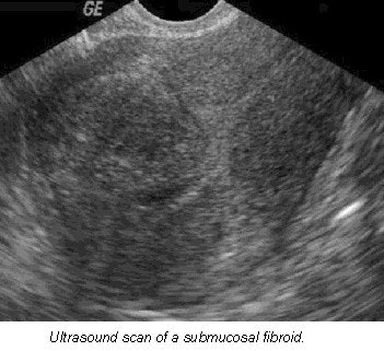
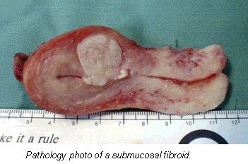
Making an accurate diagnosis of fibroids is extremely important because there are many ways of treating them. Thirty percent of women with fibroids have bleeding, particularly when the fibroids are submucosal in location.
 |
"Management options include hormone suppression, embolization, myomectomy, and finally, hysterectomy," he explained.
Lyons identified some useful clinical and ultrasound criteria that enable his department to confidently diagnose these uterine problems:
- Women with adenomyosis are usually multiparous, while women with fibroids are more likely to be nulliparous. Lyons emphasized that "a fibroid is a clearly defined, easily identified mass, whereas adenomyosis is a diffuse infiltrative process."
- Women with menorrhagia, increased volume or duration of bleeding, are far more likely to have adenomyosis than fibroids. Ultrasound features for fibroids include one or more discrete masses with a hypoechoic halo (compressed myometrium), diffuse posterior shadowing, and calcification in late stages or after pregnancy. However, cysts are uncommon.
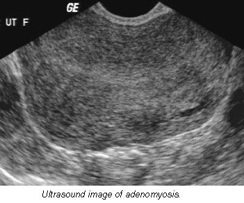
- Fibroids can be hypoechoic, isoechoic (same echogenicity as myometrium), or hyperechoic. In comparison, the characteristics of adenomyosis include ill-defined regions of mixed textural changes, asymmetric myometrial thickening, streaky shadowing posteriorly, and no calcifications.
- Myometrial cysts, often in a subendometrial location and usually less than 3 mm in diameter, are commonly associated with adenomyosis. These cysts represent dilated endometrial glands that come and go during the menstrual cycle.
"We routinely repeat the [ultrasound] study on day 19 if there is a suspicion of adenomyosis based on history or sonographic features," Lyons stated. Fibroids may undergo cystic degeneration, but this is not a common phenomenon.
 |
Also, increasing fibroid size does not make cystic degeneration more common, he said. Fibroids are seen in a number of locations. They may be subserosal, submucosal, pedunculated, or a broad ligament fibroid.
"The reason that it is important to identify where they are is that on hysteroscopy if they are pedunculated or submucosal, you can go in there, you can identify them, and begin to burn them off," he said.
 |
Color or power Doppler has been useful for vessel identification and in fibroids. According to Lyons, echogenic fibroids appear as they do because of hydropic degeneration -- not because they contain fat, as is often assumed.
Because adenomyosis is exacerbated by estrogen production, the condition regresses after menopause unless hormone replacement therapy is being administered. The only definitive treatment for adenomyosis is total hysterectomy. The drug Danazol, a gonadotropin-releasing hormone agonist, has been found to reduce uterine size and effect a reduction in the symptoms of menorrhagia and dyspareunia. Unfortunately, there is no long-term regression of adenomyosis, and recurrence of symptoms are usually documented within six months of cessation of therapy.
By Leanne McKnoultyAuntMinnie.com contributing writer
November 13, 2001
Leanne McKnoulty is an accredited medical sonographer currently lecturing in ultrasound at Monash University in Melbourne, Australia. She also is an editor at Ultrasound Review.
Copyright © 2001 AuntMinnie.com


