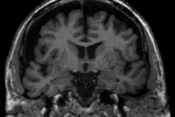
Brain imaging studies show that loneliness and social isolation cause distinct but overlapping patterns of brain changes in middle-older adults, with isolation primarily affecting temporal regions and loneliness affecting subcortical structures, both linked to increased dementia risk.
- Distinct brain patterns: Social isolation shows consistent associations with temporal brain regions like the middle temporal gyrus, while loneliness associates more with subcortical structures including the putamen and globus pallidus
- Shared neural effects: Both loneliness and social isolation affect limbic regions including the hippocampus, amygdala, thalamus, and fusiform gyrus
- Dementia connection: Several brain regions affected by both conditions are vulnerable in Alzheimer's disease and related dementias
- Biomarker findings: Both constructs linked to greater white matter hyperintensity volume and elevated C-reactive protein, an inflammatory marker
- Research gap: Prior studies often used these terms interchangeably, obscuring distinct neurobiological pathways; treating them as separate research targets is essential
MRI and PET scans reveal that social isolation and loneliness are each associated with distinct but overlapping patterns of brain change in middle-older adults, according to findings from a systematic review published October 28 in Alzheimer’s & Dementia.
A group from Deakin University in Geelong, Victoria, Australia synthesized neuroimaging and biomarker evidence examining the two social health constructs separately, with findings suggesting they affect different brain circuits and may contribute to dementia risk, noted lead author Jade Guarnera, a PhD candidate, and colleagues.
"We currently lack a strong understanding of the mechanisms that might explain this risk, particularly the underlying neurobiology," the group wrote.
Social isolation and loneliness are both recognized dementia risk factors, but prior studies frequently have used the terms interchangeably and applied measures designed for one construct when investigating the other, which obscures potentially distinct neurobiological pathways, the group wrote. Thus, the researchers aimed to synthesis current evidence and examined their neural correlates separately.
The researchers searched PubMed, PsycInfo, Medline ProQuest, and Embase for studies published between 2010 and 2025 that examined social isolation or loneliness alongside neurobiological outcomes in middle-older adult samples (mean age ≥ 50 years). Outcomes included structural and functional MRI, diffusion tensor imaging, PET (tau, beta amyloid), cerebrospinal fluid, and blood-based neuroinflammatory markers including C-reactive protein. The final review comprised 42 studies, seven examining social isolation, 26 loneliness, and nine both.
 Gray matter cortical and subcortical regions showing exclusive patterns of association with social isolation or loneliness. Highlighted regions indicate exclusive patterns of association with each construct across the literature, replicated in ≥ 2 studies. A, Social isolation. B, Loneliness.Alzheimer’s & Dementia
Both constructs shared neurobiological associations in limbic regions (the hippocampus, amygdala, thalamus, and fusiform gyrus), and each was linked to greater white matter hyperintensity volume and elevated C-reactive protein. Lastly, several of the implicated regions are known to be vulnerable in Alzheimer’s disease and related dementias, the researchers reported.
"To our knowledge, this is the first systematic review exploring the neurobiological correlates underpinning both social isolation and loneliness in middle-older adults," the authors wrote.
Ultimately, the distinct and shared neural signatures identified support the need to treat social isolation and loneliness as separate research targets, the group suggested. They encouraged future studies to use validated multi-item loneliness measures, incorporate fMRI and broader white matter metrics in older adult samples, and prioritize study designs that examine social isolation and loneliness within the same analytical framework to improve comparability.
"Moreover, the development of a multidimensional, social isolation–specific measure should be prioritized as the social health field advances," the group concluded.
The full study is available here.