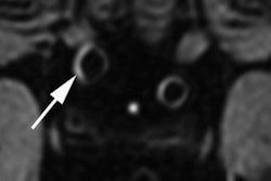
7T MRI detects significantly more multiple sclerosis lesions than conventional 3T MRI, including 27% more lesions overall and nearly double the central vein sign-positive lesions, according to a study published in Radiology.
- 7T MRI found 2,268 MS lesions compared to 1,779 with 3T MRI in the same patients
- Central vein sign-positive lesions were nearly 2.5 times higher on 7T (1,451 vs 592)
- 7T scans showed significantly higher detection across cortical (+135.7%), juxtacortical (+26.1%), periventricular (+18.8%), and infratentorial (+33.5%) regions
- 7T's superior performance is due to higher spatial resolution, improved signal-to-noise ratio, and better susceptibility-weighted contrast
- Study included 30 MS patients who underwent same-day scanning with both MRI field strengths for direct comparison
7T MRI improves multiple sclerosis (MS) lesion detection, according to findings published November 10 in Radiology.
Compared with 3T MRI, 7T scans find lesions better across characteristic intracranial regions and key imaging biomarkers, wrote a team led by Xinyao Liu from Capital Medical University in Beijing, China.
“Moreover, the proportion of central vein sign [CVS]-positive lesions was higher with 7T MRI than with 3T MRI, whereas there was no evidence of a difference in the proportion of paramagnetic rim lesions [PRLs] detected,” Liu and colleagues wrote.
MRI is key for accurately diagnosing MS. And while 7T MRI can visualize lesions more clearly than conventional 3T MRI, the researchers noted limited data that compares the two approaches head-to-head across lesion locations and imaging biomarkers.
“Consensus statements call for such studies to better define the clinical value of 7T MRI,” the researchers wrote.
 Multiple sclerosis (MS) lesions: 7T versus 3T MRI. MRI scans of cortical lesions in two participants with relapsing-remitting MS: (A) 34-year-old female and (B) 56-year-old female. For each participant, axial magnified views at 7T and 3T MRI show the lesion (white arrowheads), with insets showing the corresponding anatomic location (yellow arrowheads). The MS lesion is clearly visible at 7T MRI but difficult to visualize at 3T MRI. FLAIR = fluid-attenuated inversion recovery, T1WI = T1-weighted imaging, T2WI = T2-weighted imaging.RSNA
The Liu team compared the respective performances of 7T and 3T MRI for detecting MS lesions across characteristic intracranial regions and key imaging biomarkers. The study included 30 patients with a median age of 34.5 years who were prospectively enrolled to undergo same-day scanning with both MRI field strengths.
7T MRI visualized more MS lesions overall than 3T MRI, which included more CVS-positive lesions and PRLs.
Scanning performance of 7T, 3T MRI for MS lesion detection | |||
Measure | 3T | 7T | P value |
Lesions found | 1,779 | 2,268 | < 0.001 |
CVS-positive lesions found | 592 | 1,451 | < 0.001 |
PRLs found | 468 | 634 | < 0.001 |
7T scans also led to significantly higher detection across cortical (+135.7%), juxtacortical (+26.1%), periventricular (+18.8%), and infratentorial (+33.5%) regions (all p < 0.001).
The researchers also observed higher proportions of CVS-positive lesions on 7T MRI compared to 3T scans (64.0% vs 33.3%, p < 0.001). However, both imaging approaches led to similar proportions of PRLs (28.0% vs 26.3%, p = 0.057). Finally, the team reported excellent interrater reliability (intraclass correlation coefficient = 0.99).
7T MRI’s success can be attributed to its high spatial resolution, signal-to-noise ratio, and susceptibility-weighted contrast, the study authors highlighted.
“The former improves delineation of small lesions, especially in cortical and juxtacortical regions,” they wrote. “The latter enhances visualization of venous signals for CVS and iron-laden signals from microglia or macrophages for PRLs, thereby improving detection of both markers.”
The authors called for future guidance and clinical practice to consider the differences in the detection of MS lesions between 3T and 7T MRI.
Read the full study here.