Gadoxetic acid-enhanced MRI is transforming liver disease diagnosis by enabling earlier detection of small liver cancers and providing cellular-level assessment of liver function, with superior sensitivity compared to traditional imaging methods and integration with artificial intelligence showing promise for personalized precision medicine.
- Superior Detection: Gadoxetic acid MRI shows sensitivity of 0.82 at 2cm thresholds versus 0.53 for contrast-enhanced CT in detecting hepatocellular carcinoma
- Cellular-Level Assessment: About 50% of the contrast agent is absorbed by functional hepatocytes, allowing imaging at the cellular level rather than just morphological evaluation
- Clinical Integration: China prioritized gadoxetic acid MRI in national guidelines by 2017, with a national procurement program exceeding 1.5 million units annually
- AI-Driven Innovation: Deep learning models trained on gadoxetic acid MRI data achieve AUC scores of 0.81 for predicting microvascular invasion, outperforming CT-based approaches
- Cost-Effective: Total per-patient diagnosis and treatment costs with gadoxetic acid MRI (¥30,360) are comparable to or lower than alternative imaging modalities
A liver-specific contrast MRI agent may hold the key toward better diagnosing liver disease, suggest findings published in Quantitative Imaging in Medicine and Surgery.
Gadoxetic acid is a highly sensitive tool for detecting early small-cell liver cancer and can estimate hepatic functional reserve (HFR) and stratify risk for post-hepatectomy liver failure, wrote a team led by Yan Liu from Southern Medical University in Guangzhou, China.
“While clinical translation of advanced analytics requires rigorous, prospective multicenter validation…, combining this functional imaging agent with data-driven frameworks represents a promising frontier to eventually optimize future radiology workflows and support personalized precision hepatology,” the Liu team wrote.
Gadoxetic acid first became available for clinical use in 2011. The researchers highlighted that it has altered the direction of liver care from morphological evaluation to assessment at the cellular level.
Liu and colleagues evaluated the clinical use of gadoxetic acid-enhanced MRI (EOB-MRI) in Chinese hospitals over the past 15 years. They focused on its pharmacological benefits, advanced applications, and how the agent can be integrated with data-driven networks.
Benefits and national clinical integration
Gadoxetic acid combines extracellular distribution, which supports dynamic multiphase imaging, with hepatocyte-specific uptake. About 50% of the dose enters functional hepatocytes through the organic anion transporting polypeptide 1B3 (OATP1B3), peaking 10 to 20 minutes after injection, before excretion into bile. In the hepatobiliary phase, healthy liver appears bright while most malignant lesions, which lack functional OATP1B3, appear dark. The agent also has higher relaxivity than gadopentetate dimeglumine (Gd-DTPA; 10.7 vs. 4.8 L/mmol·s at 1.5T) and clears through both the liver and kidneys. This supports the agent’s safety in patients with organ impairment, the authors wrote.
China issued its first technical consensus in 2016, and EOB-MRI entered national guidelines by 2017. The 2024 and 2026 guidelines prioritize it for sub-centimeter hepatocellular carcinoma (HCC). A 2021 national procurement program, with annual volume above 1.5 million units, broadened access. The review contrasts China’s sensitivity-oriented strategy, which treats hepatobiliary-phase hypointensity as a primary indicator, with some guidance from LI-RADS and the American Association for the Study of Liver Diseases (AASLD).
Advanced applications
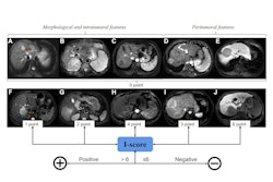
The authors also outlined several applications, including disease detection, differential diagnosis, and risk stratification, among others. The team cited meta-analysis data that showed sensitivity values of 0.82 and 0.46 at the 2 cm and 1 cm size thresholds, versus 0.53 and 0.20 for contrast-enhanced CT.
Another study found that this approach distinguished focal nodular hyperplasia from adenoma, with a pooled AUC of about 0.98, supporting an “imaging-first” approach over biopsy. Peritumoral hypointensity and multifocality showed specificities of 0.91 and 0.93 for predicting microvascular invasion.
More research suggests that relative enhancement correlates inversely with ICG-R15. And a hepatic uptake index combined with clinical markers predicted post-hepatectomy liver failure with an AUC of 0.80 versus 0.63 for a clinical score.
Finally, the 2024 guidelines recommend EOB-MRI for postoperative surveillance every three months. Total per-patient diagnosis and treatment cost after EOB-MRI (¥30,360) was slightly below that for extracellular-contrast MRI (¥31,465) and CT (¥30,803).
Integration with data-driven networks
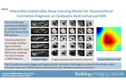
Radiomics and deep learning are converting visual signs into quantitative markers.
Reported models include a deep learning model for microvascular invasion trained on 750 HCCs from five hospitals (AUC 0.81 vs. 0.74 for CT), a CK19 nomogram (external AUC 0.74 vs. 0.60), a TP53 mutation model (retrospective AUC 0.914), and a clinical-radiomic-pathomic recurrence model (AUC 0.86). Automated segmentation tools such as nnU-Net also make way for functional liver remnant calculation in seconds on consumer-grade GPUs, the review authors wrote.
The authors argued that China has distinct advantages for this work: HBV-related disease accounts for roughly 80% of the country’s HCC burden, multimodal radiology-pathology-genomics datasets are growing, and clinician-engineer collaboration is supported by national initiatives including “Data Element×Healthcare” and “AI+ Healthcare.”
Caveats
The authors noted a gap between retrospective performance and clinical readiness, describing these frameworks as investigational tools rather than standalone decisionmakers. Challenges include data heterogeneity (transient severe motion occurs in a pooled 13.0% of exams), domain shift, and “black box” interpretability.
Next steps include explainable AI, generative AI, “digital twin” concepts, and prospective multicenter validation, the authors wrote.
“Consequently, these advancements are poised to redefine the management of liver cancer, moving toward a future where individualized, non-invasive, and high-fidelity precision medicine is accessible to all,” they added.
Read the full analysis here.