
An MRI study found that higher progesterone levels reduce the brain's ability to distinguish between different emotional situations, leading to lower mood in healthy young women. The research reveals how ovarian hormones like estradiol and progesterone directly influence brain regions responsible for emotional control and regulation.
- MRI scans showed that higher progesterone levels make it harder for the brain to distinguish between easy and difficult emotional situations
- The study included 116 naturally cycling women who performed emotional response tasks during functional MRI scans
- Brain regions affected include the lateral frontal pole and striatum, which showed reduced activity with higher progesterone
- Lower brain activity in the lateral frontal pole was associated with poorer mood during the previous week
- This research provides novel insights into how ovarian hormones regulate emotion and emotional control in women
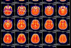
MRI reveals how the ovarian hormones estradiol and progesterone influence parts of the brain involved in emotion control in a study published September 2 in Psychological Medicine.
MRI scans showed that when progesterone levels are higher, there is a less clear distinction between “easy” and “difficult” emotional situations, corresponding to lower mood, wrote a team led by Macià Buades-Rotger, PhD, from the University of Barcelona in Spain.
“This suggests that the brain would be less able to distinguish between different emotional situations in real life,” Buades-Rotger and colleagues wrote.
Fluctuations in ovarian hormones are tied to changes in mood and emotional well-being. However, the neural mechanisms linking endocrine states and affective regulation are not well understood.
The Buades-Rotger team studied how endogenous estradiol and progesterone change brain representations behind emotion-action control in healthy young women.
The study included 116 naturally cycling women who performed an approach–avoidance task with emotional facial expressions during functional MRI.
The women completed questionnaires about their mood and period stage and gave a saliva sample to have their hormone levels measured. From there, they underwent MRI scans while conducting psychological tasks that stimulated an “easy” or “difficult” emotional response. The women were shown a happy face or an angry face and were asked to react naturally or counter intuitively. The researchers also used machine learning to analyze brain activity patterns and find out whether the participants showed an “easy” or “difficult” emotional response.
The women responded more slowly and less accurately when approaching angry faces or avoiding happy ones. This reflected control over automatic emotional tendencies, the researchers noted.
Multivoxel decoding showed that task conditions could be reliably distinguished from activation patterns in the following brain regions: lateral frontal pole, striatum, central insula, dorsal and ventral cerebellum, primary visual cortex, and periaqueductal gray.
The researchers found that progesterone levels were correlated with reduced classification accuracy in the lateral frontal pole (r = -0.26, p = 0.006) and striatum (r = -0.26, p = 0.007). And lower lateral frontal pole decodability showed associations with poorer mood during the prior week (r= -0.25, p = 0.011).
Finally, decoding accuracy in the lateral frontal pole fully mediated the indirect association between progesterone and worse mood (R2 = 7.1%).
The results provide “novel and precise insights” into the neuroendocrine modulation of affect in healthy young women, the study authors highlighted.
“While we can’t establish cause and effect from a one-time measurement, it’s exciting because we know something about progesterone that we didn’t know before,” said Buades-Rotger in a prepared statement.
Read the full study here.


.fFmgij6Hin.png?auto=compress%2Cformat&fit=crop&h=100&q=70&w=100)

.fFmgij6Hin.png?auto=compress%2Cformat&fit=crop&h=167&q=70&w=250)


