
Researchers developed an AI model that analyzes MRI scans to create detailed maps showing how different brain regions age at varying rates, revealing accelerated aging patterns in areas typically affected early by Alzheimer's disease and enabling earlier detection of cognitive decline.
- The AI model was trained on 14,748 MRI scans from cognitively normal adults and tested on over 1,900 participants from the Alzheimer's Disease Neuroimaging Initiative.
- The approach measures local brain age at the voxel level rather than relying on global brain aging measures, revealing detailed regional aging patterns previously hidden.
- Key brain structures like the hippocampus and amygdala showed significantly older local brain ages in people with mild cognitive impairment and Alzheimer's disease compared to cognitively normal adults.
- The model can enable early detection, disease monitoring, and clinical trial optimization by identifying regions with significant brain age shifts.
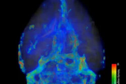
An AI model can generate detailed maps from MRI scans that highlight differences in how specific parts of the brain age, suggest findings published August 3 in Proceedings of the National Academy of Sciences.
Using MR images from people with mild cognitive impairment and Alzheimer’s disease, the AI model showed distinct patterns of accelerated aging in brain regions affected early in neurodegeneration, wrote a team led by Andrei Irimia, PhD, from the University of Southern California in Los Angeles and colleagues.
“Our approach consistently reveals spatial patterns of aging, including relatively advanced aging in frontal and temporal regions, across both typical aging and Alzheimer’s disease,” the Irimia team wrote.
Brain age mapping typically relies on global brain aging, an imaging biomarker that provides estimates of brain age. The researchers pointed out that this single summarized measure “can potentially obscure” regional patterns of cognitive vulnerability that occur prior to an Alzheimer’s disease diagnosis.
Irimia and colleagues developed their model to measure local brain age at the voxel level, 3D units that make up an MRI scan. This approach aimed to create more detailed views of structural aging in the brain.
The team trained a deep-learning neural network by using MRI scans from 14,748 cognitively normal adults ages 19 to 100 drawn from public datasets. They then tested the model using MRI scans from more than 1,900 additional participants in the Alzheimer’s Disease Neuroimaging Initiative. These participants included cognitively normal adults (n = 1,102), people with mild cognitive impairment (n = 354), and people with Alzheimer’s disease (n = 529).
The model revealed that compared to cognitively normal adults, key cortical and subcortical structures known to appear early Alzheimer’s pathology show significantly older local brain ages in both early mild cognitive impairment and Alzheimer’s disease (p < 0.05). These structures include the hippocampus, amygdala, and other deep brain regions involved in memory and cognitive processing.
The team also reported that deviations from normative regional aging are tied to cognitive performance supported by neural processes linked to those regions (p < 0.05). This finding connects anatomic aging to functional outcomes, the researchers wrote.
The study authors highlighted the potential of their AI framework to “provide anatomically interpretable measures” that can improve typical and pathological aging descriptors.
“Clinically, the approach can enable early detection and prognosis by identifying regions with significant [local brain age gap] shifts, by monitoring disease progression through [local brain age gap] tracking, and possibly even by optimizing participant selection for clinical trials based on high-risk phenotypes,” they wrote. “This scalable framework paves the way for monitoring a broad spectrum of neurodegenerative and aging-related disorders.”
The authors however added that more validation is needed, as well as more diverse clinical datasets before the model can be used in routine patient care.
Read the full study here.
.fFmgij6Hin.png?auto=compress%2Cformat&fit=crop&h=100&q=70&w=100)



.fFmgij6Hin.png?auto=compress%2Cformat&fit=crop&h=167&q=70&w=250)


