Dear AuntMinnie Member,
A new study we're highlighting in our MRI Community has disturbing implications regarding the developing problem of gadolinium retention following the administration of MRI contrast.
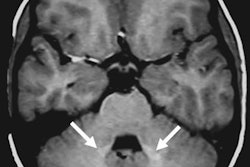
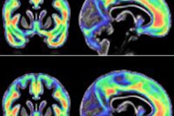
Researchers in Colorado compared a group of children who received MRI scans with a gadolinium-based contrast agent to a group of kids who had MRI scans without contrast. They found that the gadolinium group had signs of higher signal intensity in the dentate nucleus -- signs that have also been found in adults after GBCA administration.
The findings are concerning because some kids may get multiple MRI scans with gadolinium contrast over their lifetimes, and the long-term health ramifications of gadolinium retention still aren't clear. Learn more about this developing story by clicking here, or visit the community at mri.auntminnie.com.
HIMSS/SIIM on enterprise viewers
When the two most influential organizations in imaging IT speak, people listen. That's why it's important to check out our latest article on a new white paper on enterprise viewers from the Healthcare Information and Management Systems Society (HIMSS) and the Society for Imaging Informatics in Medicine (SIIM).
Interest has been rising in enterprise viewers as healthcare networks seek to unify the data from their radiology departments with nonimaging data from other sources into a single vendor-neutral archive (VNA). VNAs offer both vendor independence and easier access to disparate types of medical data.
The new HIMSS/SIIM white paper runs the gamut of considerations to keep in mind when adopting a VNA, from technical issues to security and regulatory issues. Get the rest of the story by clicking here, or visit our Imaging Informatics Community at informatics.auntminnie.com.
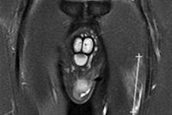
Road to Rio
As the 2016 Olympic Games open tomorrow in Rio de Janeiro, visit our AuntMinnieEurope.com sister site for a new article on how medical imaging is playing a key role in the care and treatment of the world's top athletes. The Olympic polyclinic is outfitted with a variety of imaging technology, from ultrasound to MRI. Find out how it's being used by clicking here.


![Overview of the study design. (A) The fully automated deep learning framework was developed to estimate body composition (BC) (defined as subcutaneous adipose tissue [SAT] in liters; visceral adipose tissue [VAT] in liters; skeletal muscle [SM] in liters; SM fat fraction [SMFF] as a percentage; and intramuscular adipose tissue [IMAT] in deciliters) from MRI. The fully automated framework comprised one model (model 1) to quantify different BC measures (SAT, VAT, SM, SMFF, and IMAT) as three-dimensional (3D) measures from whole-body MRI scans. The second model (model 2) was trained to identify standardized anatomic landmarks along the craniocaudal body axis (z coordinate field), which allowed for subdividing the whole-body measures into different subregions typically examined on clinical routine MRI scans (chest, abdomen, and pelvis). (B) BC was quantified from whole-body MRI in over 66,000 individuals from two large population-based cohort studies, the UK Biobank (UKB) (36,317 individuals) and the German National Cohort (NAKO) (30,291 individuals). Bar graphs show age distribution by sex and cohort. BMI = body mass index. (C) After the performance assessment of the fully automated framework, the change in BC measures, distributions, and profiles across age decades were investigated. Age-, sex-, and height-adjusted body composition reference curves were calculated and made publicly available in a web-based z-score calculator (https://circ-ml.github.io).](https://img.auntminnie.com/mindful/smg/workspaces/default/uploads/2026/05/body-comp.XgAjTfPj1W.jpg?auto=format%2Ccompress&fit=crop&h=100&q=70&w=100)


![Overview of the study design. (A) The fully automated deep learning framework was developed to estimate body composition (BC) (defined as subcutaneous adipose tissue [SAT] in liters; visceral adipose tissue [VAT] in liters; skeletal muscle [SM] in liters; SM fat fraction [SMFF] as a percentage; and intramuscular adipose tissue [IMAT] in deciliters) from MRI. The fully automated framework comprised one model (model 1) to quantify different BC measures (SAT, VAT, SM, SMFF, and IMAT) as three-dimensional (3D) measures from whole-body MRI scans. The second model (model 2) was trained to identify standardized anatomic landmarks along the craniocaudal body axis (z coordinate field), which allowed for subdividing the whole-body measures into different subregions typically examined on clinical routine MRI scans (chest, abdomen, and pelvis). (B) BC was quantified from whole-body MRI in over 66,000 individuals from two large population-based cohort studies, the UK Biobank (UKB) (36,317 individuals) and the German National Cohort (NAKO) (30,291 individuals). Bar graphs show age distribution by sex and cohort. BMI = body mass index. (C) After the performance assessment of the fully automated framework, the change in BC measures, distributions, and profiles across age decades were investigated. Age-, sex-, and height-adjusted body composition reference curves were calculated and made publicly available in a web-based z-score calculator (https://circ-ml.github.io).](https://img.auntminnie.com/mindful/smg/workspaces/default/uploads/2026/05/body-comp.XgAjTfPj1W.jpg?auto=format%2Ccompress&fit=crop&h=112&q=70&w=112)


