A new study by Canadian researchers illustrates how perfusion-based functional MRI (fMRI) with a 4-tesla arterial spin labeling (ASL) protocol can be used to measure brain activity in people with early-stage Alzheimer's disease while they perform memory tasks.
The researchers found enhanced brain activity in the bilateral prefrontal cortex and the anterior cingulate cortex in response to both memory encoding and retrieval tasks. The findings may help provide a path to an earlier Alzheimer's diagnosis, they concluded.
The study was led by Steven Beyea, PhD, scientific director of the Biomedical Translational Imaging Centre in Halifax, Nova Scotia. He presented the findings at this month's RSNA 2013 meeting.
The compensation effect
Previous studies have suggested that certain brain regions become more active to compensate for deficiencies created by Alzheimer's disease. This so-called neurocompensation response has been proposed in early Alzheimer's disease based on blood-oxygen-level dependent (BOLD) fMRI, Beyea and colleagues noted. However, few studies have evaluated this neurocompensatory response using perfusion fMRI.
"BOLD MRI, for example, can suggest a neurocompensatory response to early Alzheimer's with increased frontal activation," Beyea said. "In addition, arterial spin labeling perfusion-weighted MRI can label blood in flowing water molecules and can be more directly linked to blood supply during neural activity."
In the current study, the researchers sought to explore functional changes in the brain by scanning subjects with and without Alzheimer's disease while the subjects completed cognitive tests.
Beyea and colleagues enrolled 12 people who were diagnosed with early Alzheimer's (seven men, five women; mean age, 72.3 ± 7.9 years), and they also included 12 age-matched cognitively healthy subjects (three men, nine women; mean age, 73.7 ± 5.5 years).
ASL-fMRI scans were performed on a 4-tesla scanner with a multislice, flow-sensitive alternating inversion recovery (FAIR) protocol to visualize brain activity during two cognitive tests: one for memory encoding and one for memory retrieval.
ASL-fMRI findings
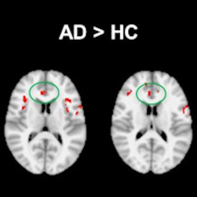
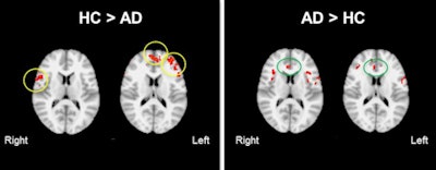
Overall, the patients with early Alzheimer's disease showed significantly greater activation in the bilateral prefrontal cortex, including the dorsolateral and ventrolateral prefrontal cortex, as well as in the anterior cingulate cortex, compared with the controls.
Brain activation patterns were most noticeably different between the Alzheimer's subjects and the healthy controls when the encoding and retrieval tasks were being performed.
The increased activity was "potentially due to the fact that increased attention was needed in these patients as they tried to use their memory," Beyea said.
 The ASL-fMR images show group differences in cerebral circulation-based functional activation in the frontal lobe between people with early Alzheimer's disease (AD) and those with cognitive healthy (HC) aging in a memory-encoding task. During memory encoding, significantly decreased activation was found in the AD group relative to healthy subjects (primarily in ventral medial and bilateral prefrontal areas). Increased activation in AD was observed mostly in the anterior cingulate cortex, suggesting that enhanced attention was required to successfully encode new information. Images courtesy of Steven Beyea, PhD.
"We see that the regions of increased perfusion are at the right prefrontal cortex, helping the Alzheimer's disease patients with the executive control of their brain, and may be due to how these patients in the early stages of Alzheimer's are compensating during the retrieval task," he said.
The findings confirm the efficacy of ultrahigh-field ASL-fMRI for detecting brain activation changes during memory encoding and retrieval tasks in patients with early Alzheimer's disease, Beyea concluded.
Patients with early Alzheimer's disease showed reduced efficiency in memory processing in specific memory networks, compared with the cognitively healthy controls.
Given the hyperactivation during the memory tasks, the Alzheimer's patients may have been exhibiting a neurocompensatory response, according to the researchers.


