VIENNA - It's early days, but the power of high-field imaging is going to push the limits of what radiologists believe is possible in breast MRI, noted Dr. Thomas Helbich, vice chair of radiology at the Medical University of Vienna, in a pre-European Congress of Radiology (ECR) meeting.
At ECR 2012, members of Helbich's department will present findings from their work with multiparametric breast imaging at 3-tesla in several scientific sessions, he noted. The first result will be to break the widely held belief that while MRI has high sensitivity for tumor detection, it has low specificity.
 3-tesla is like a Ferrari that will take us where we need to go a lot faster, according to Dr. Thomas Helbich.
"This is the mantra in radiology, and it is no longer true," he said on Tuesday during a two-day training course organized by the European Society of Breast Imaging (EUSOBI).
Helbich presented a study of 340 patients examined with dynamic contrast-enhanced MRI at 3-tesla that improved both temporal and spatial resolution and returned 99% sensitivity and 81% specificity, with a diagnostic accuracy of 93%. The higher field strength increased the signal-to-noise ratio and improved the separation of fat and water.
"It is improved at 3-tesla, but not perfect," noted Helbich, adding that low-grade ductal carcinoma in situ (DCIS) at less than 1 cm continues to elude MRI examination. "We need more and better sequences, but we are on our way and expect to achieve good results soon."
Pushing further in a standing-room-only session, Helbich introduced his group's work with what he called a multiparametric approach that combines molecular imaging with cell biology and chemistry as the foundation for novel high-field explorations using 3-tesla. This multidisciplinary approach provides a broad new approach for breast imaging, he said.
 |
| Source: 3T breast MRI: Ready to go? Dr. Thomas Helbich, EUSOBI Breast MRI Training Course 2012. |
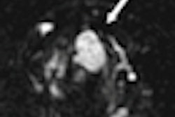
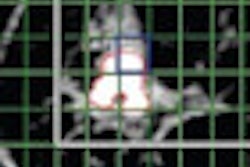
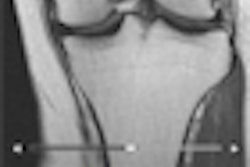
Results from a study of 112 examinations combining a contrast-enhanced MRI image for morphology and angiogenesis, a diffusion-weighted image (DWI) of cell function, and MRI-spectroscopy (MRI-S) resulted in 100% sensitivity, 89% specificity, and 96% diagnostic accuracy (see figure). Most significantly, multiparametric MRI obviates unnecessary breast biopsy in 89% of benign lesions, he noted.
A path to be explored is wider mapping of cellular function with MRI-S, which currently traces concentrations of only one metabolite, choline. Signals from phosphate and especially sodium have the potential to reveal greater telltale tumor activity.
Cancer is never a black-and-white issue, he said; it's proving to be a sophisticated adversary that is self-sustaining and requires a more intelligent solution.
Helbich displayed his enthusiasm for what he called a breakthrough approach using high-field MRI by sharing early results from the recent inclusion of PET/MRI in his group's multiparametric study. This seemed a bridge too far for the participants, half of whom admitted they had never performed an MRI breast exam.
"It sounds crazy, it may be visionary, but this is where we need to go to drive advances in the field," he said. "We are only at the beginning of 3-tesla breast imaging. It took us 20 years to fight for 1.5-tesla exams and now we have consensus, recommended indications, and robust techniques."
Breast MRI exams at 1.5 tesla are running in clinics like a good reliable engine, but 3-tesla is "a Ferrari that will take us where we need to go a lot faster," he concluded.