Dear AuntMinnie Member,
The conventional wisdom in medical imaging is that breast MRI is an invaluable tool when paired with mammography, improving breast cancer survival rates whenever it's used. But is the conventional wisdom right?
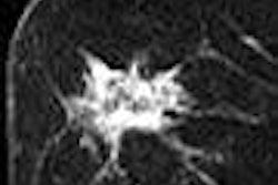
With respect to recurrent breast cancer, at least one expert isn't so sure. While breast MRI for detecting second breast cancer seems like a great idea, its value hasn't yet been proved in clinical studies, according to surgical oncologist Dr. Kimberly Van Zee of Memorial Sloan-Kettering Cancer Center and Weill Medical College of Cornell University, both in New York City.
In an article by staff writer Kate Madden Yee in our MRI Digital Community, Dr. Van Zee covers the debate over the use of MRI for recurrent breast cancer, in particular the modality's impact on patient outcomes and treatment planning.
Interestingly, she believes that the current body of research shows no benefit for MRI of recurrent breast cancers. Get the rest of the details on the story by clicking here.
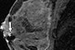
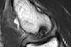
In another story on breast MRI, staff writer Shalmali Pal examines why MRI-guided breast biopsy can yield higher rates of disagreement with histologic analysis than breast biopsy using other modalities. You'll find the answer by clicking here.
Finally, a new study released today by the American Cancer Society offers further evidence that breast cancer death rates continue to fall. As in past reports, the ACS said that the cessation of hormone replacement therapy contributed to the 2% decline, but the society also said that lower mammography rates meant that fewer women are being diagnosed. Get this story by clicking here.


![Overview of the study design. (A) The fully automated deep learning framework was developed to estimate body composition (BC) (defined as subcutaneous adipose tissue [SAT] in liters; visceral adipose tissue [VAT] in liters; skeletal muscle [SM] in liters; SM fat fraction [SMFF] as a percentage; and intramuscular adipose tissue [IMAT] in deciliters) from MRI. The fully automated framework comprised one model (model 1) to quantify different BC measures (SAT, VAT, SM, SMFF, and IMAT) as three-dimensional (3D) measures from whole-body MRI scans. The second model (model 2) was trained to identify standardized anatomic landmarks along the craniocaudal body axis (z coordinate field), which allowed for subdividing the whole-body measures into different subregions typically examined on clinical routine MRI scans (chest, abdomen, and pelvis). (B) BC was quantified from whole-body MRI in over 66,000 individuals from two large population-based cohort studies, the UK Biobank (UKB) (36,317 individuals) and the German National Cohort (NAKO) (30,291 individuals). Bar graphs show age distribution by sex and cohort. BMI = body mass index. (C) After the performance assessment of the fully automated framework, the change in BC measures, distributions, and profiles across age decades were investigated. Age-, sex-, and height-adjusted body composition reference curves were calculated and made publicly available in a web-based z-score calculator (https://circ-ml.github.io).](https://img.auntminnie.com/mindful/smg/workspaces/default/uploads/2026/05/body-comp.XgAjTfPj1W.jpg?auto=format%2Ccompress&fit=crop&h=100&q=70&w=100)


![Overview of the study design. (A) The fully automated deep learning framework was developed to estimate body composition (BC) (defined as subcutaneous adipose tissue [SAT] in liters; visceral adipose tissue [VAT] in liters; skeletal muscle [SM] in liters; SM fat fraction [SMFF] as a percentage; and intramuscular adipose tissue [IMAT] in deciliters) from MRI. The fully automated framework comprised one model (model 1) to quantify different BC measures (SAT, VAT, SM, SMFF, and IMAT) as three-dimensional (3D) measures from whole-body MRI scans. The second model (model 2) was trained to identify standardized anatomic landmarks along the craniocaudal body axis (z coordinate field), which allowed for subdividing the whole-body measures into different subregions typically examined on clinical routine MRI scans (chest, abdomen, and pelvis). (B) BC was quantified from whole-body MRI in over 66,000 individuals from two large population-based cohort studies, the UK Biobank (UKB) (36,317 individuals) and the German National Cohort (NAKO) (30,291 individuals). Bar graphs show age distribution by sex and cohort. BMI = body mass index. (C) After the performance assessment of the fully automated framework, the change in BC measures, distributions, and profiles across age decades were investigated. Age-, sex-, and height-adjusted body composition reference curves were calculated and made publicly available in a web-based z-score calculator (https://circ-ml.github.io).](https://img.auntminnie.com/mindful/smg/workspaces/default/uploads/2026/05/body-comp.XgAjTfPj1W.jpg?auto=format%2Ccompress&fit=crop&h=112&q=70&w=112)


