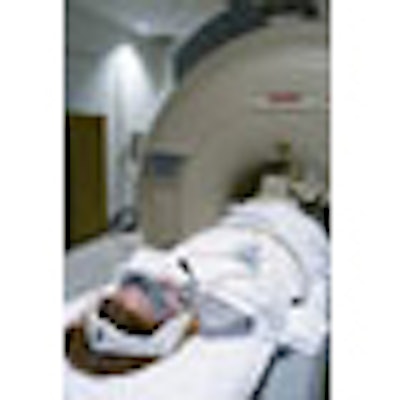
This developer of MRI-compatible patient audiovisual entertainment systems will use the 2006 RSNA conference to highlight recent enhancements to its CinemaVision product, and will introduce a new system for functional MRI (fMRI) studies.
The Northridge, CA, company will introduce a new lightweight headset for CinemaVision that integrates video display and audio systems into a single unit. The headset delivers a resolution of 240,000 pixels for each of the two displays to provide high-quality video, while the system's audio specifications include digital audio sound, passive noise-attenuation technology, and an intercom for ongoing communication between patient and technologist. The entire headset fits within an MRI coil.

For functional MRI applications, FuncLab is a new fMRI data processing system that is designed for both clinical and academic applications. It combines a functional imaging task presentation component with 3D graphics and sound capabilities with an automated data processing component, eliminating complex, time-intensive manual data analysis, according to the company.
FuncLab's data processor server attaches to the imaging facility's network, and functional and anatomic images are automatically sent in DICOM format from the MRI scanner to the processor. The system processes the data and produces brain maps of anatomy fused with functional results, with reports available over the Web through a browser-based interface.
Resonance Technology believes that FunLab will enable clinicians with no experience in fMRI to perform the procedures, creating a new revenue stream for their facility. For experienced functional labs and research institutions, the product can increase data processing speeds.
By Brian Casey
AuntMinnie.com staff writer
November 1, 2006
Copyright © 2006 AuntMinnie.com