This year has not been a stellar one for the reputation of lacrosse. Off the field, the sport, which predates the American Revolution, must contend with the Duke University rape scandal in Durham, NC. On the field, sports medicine specialists are saying that male lacrosse players are at much greater risk for potentially career-ending anterior cruciate ligament (ACL) injuries.
One possible reason that men's lacrosse is so detrimental to the ACL may be because it is a full-contact sport, according to the researchers from the Malcolm Grow Medical Center at Andrews Air Force Base in Maryland.
As a player zooms away from a looming lacrosse stick or even collides head-on with another player, he may awkwardly pivot or jump away from his opponent, resulting in excessive knee strain, which could rupture the ACL, explained lead author Dr. Anthony Beutler (American Journal of Sports Medicine, June 2006, Vol. 34:6, pp. 899-904).
While sports medicine doctors continue to look for ways to prevent ACL tears in athletes, radiologists in two recent studies have focused on imaging these injuries. In the first, U.K. investigators evaluated meniscal extrusions and associated knee joint abnormalities in young athletes. In another study, the spotlight was on meniscus extrusion and lateral meniscus root tears in ACL tears.
Associated ACL abnormalities
The meniscus plays a vital role in load transmission across the knee joint, explained Drs. Winston Rennie and David Finlay from the University Hospitals of Leicester in the U.K. They considered a meniscal extrusion to be a "displacement of 3 mm or more of the meniscus from the central margin of the tibial plateau as measured in the coronal plane" (American Journal of Roentgenology, March 2006, Vol. 186:3, pp. 791-794).
Their study group consisted of 24 professional athletes (15 soccer players and nine rugby players) who were free of knee pain and 23 age-matched active individuals. MR exams were done on a 1.5-tesla unit (Signa Horizon LX, GE Healthcare, Chalfont St. Giles, U.K.) with a knee coil. The protocol included sagittal and coronal T1-weighted spin-echo imaging and sagittal T2-weighted gradient-echo sequences. On MR, an internal meniscal signal extending to the articular surface was considered a meniscal tear.
According to the results, 48% of the athletes' knees showed evidence of meniscal extrusion with 12 lateral menisci and 15 medial menisci extruding. In this group, the authors found a significant association (79% of cases) between joint effusion and meniscal extrusion.
They also found a noteworthy connection between meniscal tears and meniscal extrusion (87% of cases), and ACL tear and subluxation (20%). In the control group, 18 knees showed meniscal extrusion but the authors found no significant associations with other knee joint abnormalities.
 |
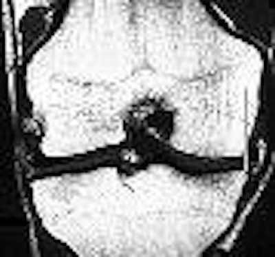
| T1-weighted coronal image of right knee at midpoint in 20-year-old asymptomatic male athlete shows medial meniscal extrusion of 5.7 mm. Meniscal extrusion is measured by distance from perpendicular line to edge of tibial plateau and edge of meniscus (white lines). Vertical tear of medial meniscus and sclerosis of subarticular medial tibial plateau with small marginal osteophyte indicating degenerative change are present. No joint effusion is shown. Rennie WJ, Finlay DBL, "Meniscal Extrusion in Young Athletes: Associated Knee Joint Abnormalities" (AJR 2006; 186:791-794). |
"All this suggests that meniscal extrusion may be an abnormal movement of the meniscus and could be a pathologic finding," Rennie and Finlay wrote. "Meniscal tears with the elimination of hoop stresses and greater or more sustained compressive forces generated by athletes within the knee could explain the significant correlation found in our study between meniscal tears and subluxation in athletes."
The duo did acknowledge that their study had certain limitations including the small number of patients, imaging performed without weight bearing, and technical constraints that would be less of an issue with an open-bore magnet.
They called for follow-up studies to evaluate whether uncorrected meniscal extrusion could lead to early degenerative changes in athletes and possibly more serious ACL injuries.
Root tears and meniscus extrusion
In the second paper, Dr. Jeffrey Brody and colleagues investigated the prevalence of posterior lateral meniscus root tears (LMRTs) in patients with an ACL tear. They also looked at the association between LMRTs with lateral meniscus extrusion and other ligament injuries.
"There is comparatively little written about tears at the anterior or posterior attachment of the meniscus to the central tibia, which is referred to as the root of the meniscus," wrote Brody's group from Brown Medical School and Rhode Island Hospital, both in Providence. "In our study, we found that LMRTs were more prevalent than MMRTs (medial meniscus root tears) in patients with ACL tears and that lateral meniscus extrusion was associated with root, complex, and deep radial tears" (Radiology, June 2006, Vol. 239:3, pp. 805-810).
This was a retrospective review for 264 MR exams done on a 1-tesla whole-body system (Magnetom Harmony, Siemens Medical Solutions, Malvern, PA). Patients were in the supine position during the exam with the knee in a standard extremity coil. The MR protocol included a T2-weighted transverse fast spin-echo sequence and sagittal T2-weighted imaging.
The MR images of the knee were reviewed for the following:
- Menicus root tears
- Other meniscus tears
- Meniscus extrusion
- Presence of meniscofemoral ligaments (MFL)
- Medial collateral ligament tears
- Bone marrow contusions
"Meniscal root tear was defined as distortion and increased signal intensity within the meniscal root that extended to the articular surface ... on MR images," the authors stated. Brody reviewed knee arthroscopy data after imaging.
 |
| LMRT and meniscus extrusion in a 20-year-old man with ACL tear. Above, coronal posterior and midknee (below) intermediate-weighted (4083/17) fast SE MR images of the right knee show ACL tear (arrowhead), LMRT (black arrow), and normal medial meniscus root (thin white arrow). The distance between the two vertical lines in image below indicates meniscus extrusion of 3.7 mm (thick white arrow). |
 |
| Figure 2a,b. Brody JM, Lin HM, Hulstyn MJ, Tung GA. Lateral Meniscus Root Tear and Meniscus Extrusion with Anterior Cruciate Ligament Tear. Radiology 2006;239:805-810. |
Among the 264 studies, 34 meniscus root tears were identified in 33 patients. LMRTs were found in 26 (9.8%) of 264 studies and MMRTs occurred in eight (3%). Lateral meniscus extrusion was present in 23% of those 26 studies and at least one MFL was seen in 81% of the cases. Extrusion of the meniscus was seen in 88% of the MMRT cases.
In other results, 82% of the 35 meniscus root tears were associated with additional meniscal tears. But the authors found no significant difference between MMRTs and LMRTs for bone marrow contusion or medial collateral ligament tears. Finally, they determined that the absence of the MFL was associated with lateral meniscus extrusion in LMRT.
The group came to several conclusions based on their research: LMRTs occurred with ACL tears and was associated with lateral extrusion of the lateral meniscus. Also, the absence of the MFL was more common when the lateral root was torn and the meniscus was extruded.
By Shalmali Pal
AuntMinnie.com staff writer
June 30, 2006
Image at top courtesy of U.S. Lacrosse.
Related Reading
3D CT measures accurately for total knee arthroplasty, May 31, 2006
Excellent long-term results from endoscopic ACL reconstruction, May 15, 2006
Return to sport after ACL tear questionable, May 24, 2005
Copyright © 2006 AuntMinnie.com