A deep learning (DL)-based brain metastasis detection model (BMDM) can substantially improve both the speed and the accuracy with which radiologists identify brain metastases on MRI scans, according to a study published March 13 in Academic Radiology.
"[Our] results demonstrated that [the model] significantly enhanced time efficiency and diagnostic performance for brain metastases detection, providing clinical benefits," wrote a team led by Meiqi Hua of the Affiliated Hospital of Hebei University/School of Clinical Medicine in Baoding, China.
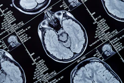
Brain metastases affect 20% to 40% of cancer patients and carry a poor prognosis, with median survival measured in months, the researchers noted -- which underscores the importance of early and accurate detection with brain MRI to effectively plan treatment and positively influence patient outcomes.
Yet conventional MRI interpretation can be time-consuming and dependent on clinician experience, the team explained. To address the problem, the group developed and validated a deep learning-based brain metastasis detection model (BMDM). The model was built on a multiscale feature pyramid network architecture designed to detect lesions of varying sizes and locations within the brain.
The group trained the model on data from 950 patients and validated it using data from an additional, independent cohort of 423 patients. It also tested three reading modes: radiologists only (four individuals with at least three years of experience and six with more than three years of experience), BMDM only, and radiologists assisted by the BMDM.
The researchers found the following:
- Use of the BMDM as a reading aid slashed average image interpretation time by 31%, from 144 seconds to 100 seconds per case.
- The model improved diagnostic accuracy, raising the area under the receiver operating curve (AUROC) from 0.84 to 0.95.
- The BMDM boosted lesion level sensitivity from 68% to 92%. This sensitivity improvement translated to a 33.4% increase in identifying micrometastases 3 mm or smaller, and a 43% increase in identifying lesions located in anatomically complex regions of the brain, the group noted.
Finally, the authors also reported that use of the algorithm benefited those radiologists with less experience: Junior readers showed a 24.6% sensitivity improvement, compared with an improvement of 22% for experienced radiologists.
Access the full study here.