Crucial to life, fast-moving and inherently variable in motion, the heart has resisted attempts to image it crisply in every modality. Not for nothing has it been called the Holy Grail of medical imaging.
Cardiac MRI in particular has struggled with the effects of cardiac motion, though it has improved tremendously in recent years with the development of high field strength clinical magnets, high-performance gradient hardware, and ultrafast pulse-sequence technologies.
But owing to beat-to-beat variation in heart motion, state-of-the-art imaging methods often suffer from a lack of robustness and repeatability, even in healthy subjects.
"Coronary MR users have long been plagued by sometimes getting poor results even when respiratory gating and ECG triggering seem to be working well," wrote Christine Lorenz, Ph.D., of Siemens Medical Solutions research in an e-mail to AuntMinnie.com.
A new study from Malvern, PA-based Siemens and John Hopkins University in Baltimore proposes an adaptive trigger delay method to compensate for beat-to-beat variations in coronary motion.
"We hypothesized that a large part of the problem could be due to variability in heart motion from beat to beat, even in healthy volunteers," Lorenz wrote. "We developed a method that tracks the motion of the coronaries in real-time and then uses that motion to derive an 'optimal' position in the cardiac cycle for acquisition of high-resolution coronary images -- this we call the adaptive trigger delay."
Recent subject-specific approaches are limited by the motion model built initially during the prescan planning, explained Maneesh Dewan; Gregory Hager, Ph.D.; Steven Shea, Ph.D.; and Lorenz in a poster presented at the 2006 International Society for Magnetic Resonance in Medicine (ISMRM) conference in Seattle. "The model built offline is not updated online during acquisition to account for variability in coronary motion," they wrote.
Current models also rely on a priori knowledge of the primary direction of motion of the coronary arteries with regard to heart orientation.
The method begins with detection of the R wave in the ECG signal. Free-breathing real-time MR images are acquired in preselected views, and coronary locations are tracked in the real-time images. These data are used to predict the triggering delay and data acquisition window. Then high-resolution data are acquired during the predicted acquisition window. The process then reverts to real-time imaging, the acquired data are corrected retrospectively, and the cycle begins again.
In the study of five healthy volunteers, true-FISP MR images were acquired in four-chamber short-axis and coronal views at TR/TE/FL 1.7-2.7/0.85-1.35/50-55. In-plane resolution was 1.5-2.4 mm, slice thickness was 8 mm on a 1.5-tesla Espree scanner (Siemens Medical Solutions). Approximately 750 frames ware acquired during free breathing at 10-15 frames/sec in the volunteers, the team wrote.
To track and then extract the coronary motion, fat-filled grooves where the coronary arteries sit were used as a surrogate for tracking coronary motion. "The tracking algorithm uses a variation of the sum of squared differences with multiple templates," the team wrote. The tracked motion data are filtered with Savitzky-Golay and Gaussian filters to remove noise.
"In this preliminary study, we used motion patterns extracted from the images of volunteer subjects, and experimented with the effect on image quality of using different image reconstruction schemes," Lorenz explained.
 |
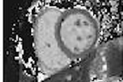
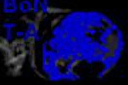
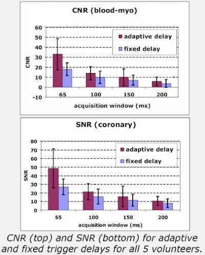
The images above show the results in a synthetic image derived from the real motion data of two subjects using the adaptive trigger delay. Images below shows results from using an optimal fixed trigger delay. Motion artifact was significantly reduced in the images with adaptive trigger delay compared to those acquired with a fixed delay, even for relatively long acquisition windows. The resulting signal-to-noise ratio (SNR) and contrast-to-noise ratio (CNR) were also improved.
 |
The use of adaptive trigger delays for each heartbeat may improve overall motion compensation, especially in cases where heart rate variability is high, the authors concluded. The subject-specific approach is feasible, "and has potential advantages over existing methods by reducing assumptions regarding motion of the coronary arteries," they wrote.
The next goal will be to integrate the tracking method into the MR scanner to further evaluate the approach. Eventually, the method could potentially be extended to other cardiac structures, such as valves, that undergo complex motion, Dewan and colleagues wrote.
By Eric Barnes
AuntMinnie.com staff writer
June 6, 2006
All charts and images courtesy of Siemens Medical Solutions.
Related Reading
High osmolarity agent beats gadolinium in cardiac MR, May 30, 2006
Perfusion MRI: A multifaceted modality for myocardial ischemia, May 8, 2006
MR angiography offers greater detail in vascular imaging, March 10, 2006
MRI immediately post MI yields important prognostic clues, December 1, 2005
Copyright © 2006 AuntMinnie.com