When it comes to forging new imaging tools, Northern California's Stanford University is a virtual factory. The school's substantial resources, talented gene pool, and unusually close ties between radiology and electrical engineering have all nurtured its success in radiology research over the years, notably in 3-D imaging, image-guided surgery, and MRI.
Recently, for example, researchers at Stanford's Magnetic Resonance Systems Research Laboratory fine-tuned a technique known as real-time color flow MRI. Originally developed five years ago to study cardiac motion and blood flow, the technique has now been modified to track the slower velocities seen in skeletal muscle motion -- velocities ranging from 0.2 to 0.5 meters per second.
At the November 2000 RSNA meeting, Dr. Garry Gold described the methods his team has used to visualize muscle velocities, calling them "a paradigm shift from static MRI" that could improve the diagnosis and treatment of patients with many kinds of motion difficulties.
"Dynamic imaging techniques have made it possible to study muscle motion noninvasively," Gold said. "Cine-PC [phase-contrast] MRI, which has been used in past, requires repeated cycles of motion to acquire images representing one motion cycle. This is simply different. This is imaging in real-time like ultrasound.... I encourage everybody to think about ways in which this technology might modify your practice."
Five normal volunteers were used for the study, in which spiral phase-contrast data is acquired continuously, using real-time gridding and phase differences to compute density and velocity maps. The maps are then displayed by means of a color overlay similar to that used in ultrasound.
Gold and colleagues Scott Delp, Krishna Nayak, and Deanna Askakawa used a flexible gray-scale coil to acquire images of biceps and triceps in motion. The trajectory covered a full 90º of flexion in approximately 35 seconds. Images were acquired with an in-plane resolution of 2.4 mm and a 12-cm field of view, and resolution was relatively coarse at 160 x 160 pixels.
"We developed a real-time phase-contrast spiral MRI sequence using a spiral k-space trajectory velocity encoding of 0.2 meters per second," Gold said.
Data was acquired rapidly through the trajectory to minimize susceptibility to overflow and motion artifacts. Images were acquired at the rate of 6 images per second, then interpolated to achieve a display rate of up to 18 images per second, which is sufficient to see real-time motion, he said.
"We acquire one full case-based set of data with the flow-encoding gradient off, and then a second one with the flow-encoding gradient on. You do this again with a slightly different spiral trajectory and then a third time, and these three sets represent one complete image."
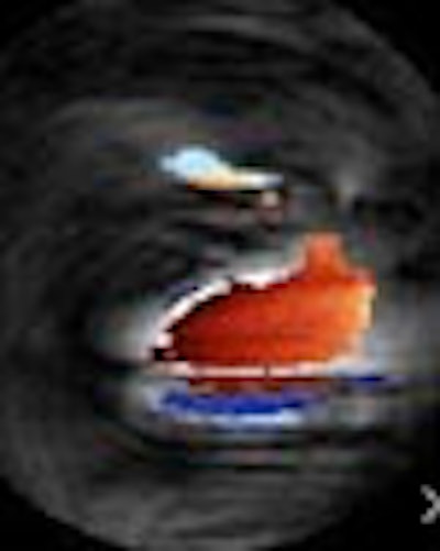
The resulting image is used to reconstruct a magnitude image, then both images are used to create a phase-difference image. Finally, the phase-difference image is used to create density and velocity images, which are overlaid as a color map on the routine gray-scale image to enable visualization of the biceps and triceps velocities.

Image (above) and QuickTime movie (below) show an axial slice through the subject's upper arm, with the biceps muscle highlighted in red (top) moving towards the viewer, and the triceps muscle in blue (bottom) moving away from the viewer. All images courtesy of Garry Gold, MD, and Stanford University Magnetic Resonance Systems Research Laboratory © 2000.
|
|
|
"Our velocity measurements are consistent with those that have been measured in the past in cine-PC MRI," Gold said. "We see real-time muscle contraction patterns in all volunteers, and antagonistic muscles moving in opposite directions with each motion cycle."
The technique could yield new information about biomechanics, he said.
"One of the many advantages of the real-time system is that it allows us to fly through the patient in any orientation, and prescribe the point that we want by dropping down to, say, an elbow weak point that happens to be axial to the biceps in the direction of the patient's spine."
Real-time MRI is both promising and feasible for studying muscle motion and velocities in real-time, Gold concluded. It can be used to study maximum force velocity patterns, to assess the efficacy of tendon transfer surgery in patients with cerebral palsy, and to evaluate patients who have difficulty with repeatable motion.
By Eric BarnesAuntMinnie.com staff writer
February 2, 2001
Click here to post your comments about this story. Please include the headline of the article in your message.
Copyright © 2001 AuntMinnie.com