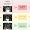
GLP-1 receptor agonists such as semaglutide and tirzepatide have little effect on FDG-PET imaging findings, according to research presented at ARRS 2026 in Pittsburgh.
The finding is from a retrospective review of 126 patients who underwent FDG-PET/CT between July 2024 and February 2025 while receiving GLP-1 agonist therapy for diabetes or weight management, noted lead author Anna Eshghi, DO, of the University of Arkansas, and colleagues.
“FDG-PET/CT can be performed in patients on GLP-1 agonist therapy, regardless of the timing of the last injection, without significantly affecting FDG biodistribution,” the group wrote.
The widespread adoption of GLP-1 agonists has raised questions for nuclear medicine departments about whether the drugs alter FDG radiotracer uptake in ways that could complicate image interpretation. FDG is a glucose analog that competes with native glucose for cellular uptake, and drugs that significantly alter glucose metabolism could theoretically shift tracer distribution across tissues, the researchers explained.
Yet previous work has not established clear guidance on whether patients should interrupt therapy before scanning or whether scan timing relative to the last dose matters, the authors noted.
Of the 126 subjects in the study, 36 patients had a prior FDG-PET/CT before starting GLP-1 therapy, allowing for paired comparison. An experienced nuclear medicine reader measured mean standardized uptake values (SUVmean), maximum SUV, and peak SUV in the blood pool, liver, gluteal muscle, and abdominal subcutaneous fat, as well as whole brain segmentation on both pre- and post-therapy scans. The researchers then compared pre- and post-therapy SUVs, and assessed associations with blood glucose, BMI, diabetes, time since last GLP-1 dose (<6 days versus = 6 days), and drug type.
According to the results, GLP-1 dosing before PET/CT ranged from zero to 10 days. Mean blood glucose among the patients decreased from 124.8 mg/dL pretherapy to 111.1 mg/dL posttherapy; BMI decreased from 37.5 to 34.7. The only statistically significant SUV change was in whole-brain SUVmean (p = 0.0274), which correlated with blood glucose level. Gluteal muscle SUVmax (p = 0.0497) and subcutaneous fat SUVpeak (p = 0.0139) showed significant correlations with BMI change.
“No significant differences in FDG uptake were observed in the skeletal muscle, liver, blood pool, or abdominal fat following GLP-1 agonist therapy,” the group wrote.
Future prospective studies with larger sample sizes and longer follow-up could further characterize any subtle longitudinal SUV changes as patients remain on GLP-1 therapy, the researchers concluded.