PET scans have revealed that higher levels of brain tau protein are associated with faster cognitive decline in women with Alzheimer’s disease than in men, a team in Boston has reported.
In a group of 1,007 cognitively unimpaired adults who were tracked over eight years, women initially outperformed men at low tau burden levels, yet the advantage diminished as tau levels increased, resulting in greater cognitive decline, noted Annie Li, MD, of Massachusetts General Hospital, and colleagues.
“Our findings highlight the importance of incorporating sex-specific tau pathology patterns into precision medicine approaches to optimize the timing and targeting of disease-modifying therapies,” the group wrote.
The build-up of tau neurofibrillary tangles in the brain is a key driver of Alzheimer’s disease symptoms, along with beta-amyloid deposits. The prevalence of the disease is significantly higher for women, with both postmortem findings and PET studies reporting greater regional tau aggregation in women compared with men, the researchers explained.
Yet few studies have investigated the sex-specific role of tau in predicting cognitive decline, particularly in the preclinical phase of the disease, they noted. To bridge the gap, the group conducted a large longitudinal meta-analysis study of more than 1,007 participants that included tau PET and amyloid PET scans and over half a decade of cognitive assessment data from three previous studies.
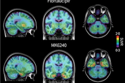
Participants ranged in age from 46 to 93, 648 (64%) were women, and 367 (39%) were carriers of the apolipoprotein E (APOE) ε4 susceptibility gene. All patients underwent beta-amyloid PET scans either with F-18 flortaucipir or F-18 MK-6240 PET radiotracer, with findings based on standardized uptake value ratios (SUVRs) of the tracers in specific brain areas of interest.
According to the results, higher temporal tau SUVR was associated with significantly faster cognitive decline in women than men, based on their performances over an average of 8.6 years on standard cognitive tests.
Specifically, women had higher tau than men across four regions: the parahippocampal gyrus (β = −0.1, p = 0.02), the fusiform gyrus (β = −0.08, p < 0.01), the inferior temporal gyrus (β = −0.09, p = 0.02), and the middle temporal gyrus (β = −0.06, p = 0.01), which are regions associated with early and later stage tau spread, the researchers reported.
“Across three longitudinal cohorts, we found that women with higher temporal tau declined faster than men,” the group wrote.
To date, there has been only one other smaller study that examined how tau burden differentially impacts rates of cognitive decline in men and women in preclinical Alzheimer’s disease, the authors noted. The strength of this study includes its large, multicohort design across three well-characterized independent cohorts with long-term follow-up, they wrote.
“Future work should identify mechanisms underlying women’s greater vulnerability to tau-related cognitive decline and examine whether similar differences occur with tau accumulation,” Li and colleagues concluded.
The full study is available here.