An AI algorithm designed for brain PET imaging found a glioblastoma in a patient that had gone undetected by physicians, according to a case reported February 15 in the Journal of Nuclear Medicine.
“This incidental finding highlights the potential of AI-based decision support for patient management in terms of diagnostic and treatment planning based on amino acid PET,” noted lead author Philipp Lohmann, PhD, of Aachen University in Aachen, and colleagues.
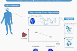
In brief, the deep learning-based AI model (called “JuST_BrainPET”) is designed to automatically segment metabolic tumor volume (MTV) from surrounding healthy tissue on brain PET imaging, a key step in medical imaging analysis, the researchers explained. The group previously described developing the AI model in an article published last year.
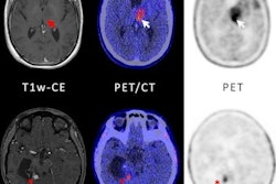
In this case, a 43-year-old man underwent an MRI scan that showed no contrast enhancement, yet hyperintensities were apparent in the patient’s left thalamus and frontoparietal region. Thus, the clinicians performed an additional PET scan with an amino acid radiotracer (F-18 FET) for further diagnosis of a suspected glioma.
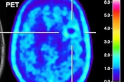
 Baseline scan (A), segmentation results (B), and follow-up scan (C) from patient with molecular glioblastoma. Baseline MRI showed FLAIR hyperintensities in left thalamus and frontoparietal region (white arrowheads). In contrast to expert segmentation, in which only left thalamic region showed slightly increased uptake (red contour), AI algorithm identified additional frontoparietal lesion on baseline PET that subsequently progressed to contrast-enhancing and metabolically active lesion (red arrowheads). Image courtesy of the Journal of Nuclear Medicine.
An analysis of the PET scan by an experienced nuclear medicine physician showed an increase in F-18 FET radiotracer uptake that revealed a potential tumor in the patient’s left thalamic region, the authors noted.
Subsequently, the researchers tested their JuST_BrainPET tool on the baseline F-18 FET-PET image.
“Surprisingly, a second lesion in the frontoparietal region, not segmented by the expert, was identified by the AI algorithm,” the group wrote.
Moreover, on follow-up imaging four months later, the patient’s left thalamic lesion showed no progression, while the additional frontoparietal lesion identified by the AI model had progressed to become a small contrast-enhancing and metabolically active tumor, the group wrote.
“The AI tool correctly predicted a pathologic process at an early disease stage and could have potentially influenced diagnostic and treatment decisions, such as biopsy guidance and target volume definition for radiotherapy,” the group concluded.
A link to the full case report can be found here.