A group in Germany has developed an AI model for PET imaging that could help clinicians determine whether patients are responding to brain tumor treatments, according to a study published August 10 in the Journal of Nuclear Medicine.
Researchers led by Robin Gutsche, PhD, of Aachen University in Aachen, demonstrated that a deep learning-based AI model can automatically segment metabolic tumor volume (MTV) on brain PET imaging. They suggested the approach could replace the need for such image preprocessing by clinicians.
"Our deep learning-based F-18 FET PET segmentation allows a reliable, robust, and fully automated evaluation of MTV in patients with brain tumors," the group wrote.
Evaluation of MTV changes using PET scans with amino acid radiotracers such as F-18 fluoroethyl tyrosine (FET) is an important tool for assessing treatment response in brain tumor patients. MTV is usually determined by manual or semiautomatic delineation, however, which is laborious and may also be prone to variability by clinicians, the researchers noted.
In this study, the group aimed to develop a method for automated MTV segmentation and to evaluate its performance for response assessment in patients with gliomas.
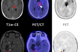
The researchers included a total of 699 F-18 FET-PET scans from 555 brain tumor patients that were previously acquired at initial diagnosis or during follow-up visits. Experienced nuclear-medicine physicians first segmented MTV in the images, which included lesions with low to high F-18 FET radiotracer uptake.
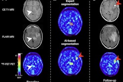
 A visual abstract. Image courtesy of the Journal of Nuclear Medicine.
Next, the researchers used 476 of these images to train an AI neural network (called "no new U-Net") to segment MTV. To test the model's performance, the group used an imaging dataset of 223 scans from 156 patients.
According to the results, out of 205 lesions with increased F-18 FET uptake in the dataset, the model correctly identified 189. Importantly, none of the anatomic regions that showed a physiologically increased uptake of F-18 FET radiotracer, such as in the superior sagittal sinus, were considered to be tumors by the network, the authors noted.
Overall, the model achieved a mean F1 score of 92%, a sensitivity of 93%, and a positive predictive value of 95% for lesion detection, according to the findings.
"This finding highlights the value of the network for improvement and automatization of clinical decision-making based on the volumetric evaluation of amino acid PET," the researchers wrote.
The authors noted that an advantage of the model is that it is designed to fully automate 3D segmentation using a single F-18 FET-PET scan and a conventional graphics processing unit in less than two minutes without preprocessing. This suggests it could be highly useful in clinical practice, they wrote.
"The main finding of our study is that our deep learning-based neural network allows reliable and fully automated detection and 3D segmentation of brain tumors investigated by F-18 FET PET," the group concluded.
A link to the full article can be found here.