PET/CT scans have the potential to predict brain metastasis in melanoma patients, according to a study published December 26 in Cancers.
In a retrospective study, a team at the University Hospital Salzburg in Austria studied baseline F-18 FDG-PET/CT imaging parameters in patients with melanoma and found they were associated with the development of metastatic brain tumors.
“In melanoma patients, identification of those who are at higher risk of brain metastasis occurrence can be crucial as subsequent surveillance and even prophylactic treatment options can be provided,” wrote lead author and nuclear medicine physician Forough Kalantari, MD.
Malignant melanoma is an aggressive, highly metastatic cancer and has the highest mortality rate among all skin-related malignancies, the authors noted. Moreover, melanoma is the third most common metastatic tumor to the brain and accounts for roughly 10% of all developed brain metastases, they added.
While F-18 FDG-PET/CT is an accurate imaging modality for detecting melanoma metastases, data on its baseline prognostic value in these patients are scarce, they added.
To that end, the group investigated the prognostic value of F-18 FDG-PET/CT in predicting the occurrence of brain metastases in 159 consecutive patients with proven melanoma who underwent imaging at their hospital in Salzburg between 2008 and 2021.
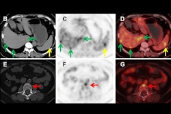
 A 49-year-old patient with primary clinical stage III melanoma underwent F-18 FDG-PET. The primary tumor Breslow thickness was 6.5 mm. On initial F-18 FDG-PET images, this patient had a high SUVmax (52), resulting in limited survival with the occurrence of brain metastasis in the first year of follow-up (horizontal arrow). Notably, during the course of the treatment, the patient underwent surgical removal of the primary tumor as well as lymph node dissection, followed by neoadjuvant radiotherapy and immunotherapy. Image courtesy of Cancers.
All patients underwent F-18 FDG-PET/CT for initial staging, as well as serial imaging follow-ups for diagnosing brain metastasis. The researchers culled baseline parameters from the PET/CT imaging and correlated the parameters with the occurrence of brain metastases using risk regression models. The median follow-up was six years.
According to the findings, baseline peak standardized uptake values (pSULpeak) – a parameter representing the highest FDG radiotracer activity in target tumors – were associated with earlier brain tumors.
For example, if a patient had a high pSULpeak value of 19, then the risk of brain metastasis development was 80% after approximately 53 months, and with a pSULpeak value of 26, the patient had an 80% risk after about 39 months, they wrote.
“As the baseline pSULpeak increased, the likelihood of the patient developing brain metastases rose and occurred at an earlier time,” the group wrote.
Ultimately, the findings could help clinicians identify those patients who could benefit more from regular follow-ups or who may require additional prophylactic treatments, the group noted.
“Our results may suggest that in addition to clinical and pathological characteristics of tumors, F-18 FDG-PET/CT findings can be of value for prognostic risk stratification considering the occurrence of brain metastasis,” the researchers concluded.
The full study can be found here.