PET/CT has significant advantages over conventional methods for lesion detection and staging in patients with natural killer/T-cell lymphoma, according to a study published December 18 in Heliyon.
In a retrospective analysis, a team in Taiyuan, China, found that F-18 FDG-PET/CT was superior for analyzing imaging manifestations of NK/T-cell lymphoma and suggested that the scans can improve treatment plans for patients.
“F-18 FDG-PET/CT scanning is crucial in identifying tumor lesions, determining staging, and devising treatment strategies for individuals diagnosed with NK/T-cell lymphoma,” wrote lead author Huixia Geng, MD, of the Third Hospital of Shanxi Medical University.
NK/T-cell lymphoma originates from mature T cells and NK cells; while the disease has a low incidence, it progresses rapidly and patients have low survival outcomes, the authors explained.
Previous studies have shown that F-18 FDG-PET/CT is key in determining the staging, response assessment, and prognostic evaluation of diffuse large B-cell lymphoma, Hodgkin's lymphoma, and follicular lymphoma, yet has not been established in patients with NK/T-cell lymphoma, they added.
To that end, the group analyzed PET/CT imaging characteristics of 38 patients with a primary diagnosis of NK/T-cell lymphoma and compared the approach with conventional methods: namely, physical examination, CT with intravenous contrast, MRI, biopsies from primary sites, and bone marrow exams.
According to the analysis, the patients had 219 lesions in total (including 81 nodal lesions and 138 extranodal lesions) that tested positive for malignancy. PET/CT outperformed conventional methods in detecting malignant lesions by a significant margin, detecting 79 (98.8 %) extranodal lesions compared to 45 (56.3 %).
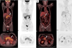
 A 50-year-old man with recently diagnosed nasal-type NK/T-cell lymphoma underwent an F-18 FDG-PET/CT scan (A–D) for initial staging. Maximum-intensity-projection image (A) shows hypermetabolic lesions in both ethmoid sinus (thin arrow), both cervical lymphatic chains (arrowhead), left upper lung (thick arrow), liver, and spleen. Transaxial images show an F-18 FDG-avid mass in both ethmoid sinus (B). The scan shows intense F-18 FDG radiotracer uptake in left upper lung and uptake in the liver and spleen (CT showed no lesions), suggesting malignancy (C–D). Finally, after the PET/CT scan was done, the patient’s staging was changed from II to IV.Image courtesy of Heliyon
In addition, the clinical staging of 15.7 % of patients (6/38 cases) was restaged to varying degrees after PET/CT findings, and PET/CT results helped in changing the treatment plan for 10.5 % (4/38) of patients, the group wrote.
Specifically, in two cases, treatment was changed from only radiotherapy to radiotherapy combined with chemotherapy, and in two cases, it was changed from radiotherapy combined with chemotherapy to only chemotherapy due to upstaging. PET/CT scans changed the radiotherapy target area in three patients, the researchers added.
“Our results suggest that PET/CT has a significant advantage over [conventional methods] for lesion detection and staging of extranodal NK/T-cell lymphomas,” the group wrote.
Ultimately, the advantage of PET/CT is that it combines both structural imaging and functional metabolic information, the authors noted. This noninvasive hybrid approach allows clinicians to analyze the morphology, location, and invasion of lesions in a single examination, compared to a battery of conventional methods, according to the authors.
“The findings of our study on NK/T-cell lymphomas highlight the significance of PET/CT in lesion detection and initial staging,” the group concluded.
The full article can be found here.