F-18 FAPI-PET/CT may be superior to F-18 FDG-PET/CT for diagnosing primary and metastatic lung cancer, according to a study published August 8 in Radiology.
Researchers led by Yuchun Wei, MD, PhD, of Shandong University Cancer Center in Jinan, China, analyzed imaging from lung cancer patients in a clinical trial who underwent both scans. Their findings suggest that F-18 FAPI-PET/CT is better than F-18 FDG-PET/CT, especially in detecting cancer that has spread to lymph nodes, brain, bone, and liver.
"F-18 FAPI-PET/CT may help improve the accuracy of lung cancer staging," the group wrote.
Fibroblast activation protein inhibitor (FAPI) is a ligand designed to bind to fibroblast cells involved in the growth of tumors. FAPI can be combined with radioisotopes such as gallium-68 (Ga-68) or F-18 to form PET radiotracers that reveal this activity. While previous studies have investigated the use of Ga-68 FAPI-PET/CT in patients with lung cancer, data on the use of F-18 FAPI-PET in these patients is scarce, they noted.
"No studies of the value of F-18 FAPI PET/CT for the diagnosis and staging of lung cancer have been reported," the researchers wrote.
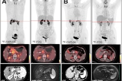
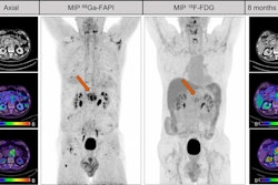
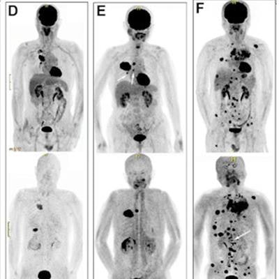
 (A-F) Representative F-18 FDG and F-18 FAPI-PET/CT scans for six study participants with lung cancer. F-18 FAPI and F-18 FDG-PET/CT images show that uptake occurred in granulomatous lymph nodes (LNs) (false-positive nodes; participant number 4, A); F-18 FAPI- PET/CT outperformed F-18 FDG-PET/CT in detecting liver metastases (participant number 21, B) and mediastinal LN metastases (participants number 22, C; 30, D; and 54, E); and F-18 FAPI- PET/CT outperformed F-18 FDG-PET/CT in detecting bone metastases (participant number 27, F). White arrows indicate foci. Image and caption courtesy of the RSNA.
To address this gap, the researchers analyzed imaging from 68 lung cancer patients in a clinical trial who underwent imaging using both F-18 FAPI-PET/CT and F-18 FDG-PET/CT, the latter of which is currently the most widely used approach. Exams were performed at intervals of more than 20 hours and within seven days of each other.
In total, 548 lesions were found. According to the analysis, the mean tumor-to-background ratio for F-18 FAPI radiotracer uptake was lower than that of F-18 FDG at the site of primary disease, yet higher at sites of lymph node spread (7.5 vs. 5.9) and bone metastases (8.6 vs. 4.3). Overall, FAPI-PET/CT showed a higher sensitivity, specificity, accuracy, and negative predictive value, the authors reported.
| F-18 FAPI-PET/CT versus F-18 FDG-PET/CT for diagnosing lung cancer | ||
| Measure | F-18 FDG-PET/CT | F-18 FAPI-PET/CT |
| Sensitivity | 97% | 99% |
| Specificity | 79% | 93% |
| Accuracy | 85% | 97% |
| Negative predictive value | 70% | 97% |
No evidence of a difference was found for positive predictive value (98% versus 92%), the group added.
Further studies using FAPI labeled with F-18 are warranted, the authors urged. F-18 has a longer half-life (110 minutes) and a high positron energy compared with other radioisotopes used to make FAPI radiotracers (specifically, Ga-68), and this could translate to more patients being screened, they noted.
"A multicenter study with a larger study sample is necessary to further confirm the observations in this study," the authors concluded.
A link to the full article can be found here.