Fibroblast activation protein inhibitor (FAPI)-PET imaging shows promise in visualizing different nonischemic cardiomyopathies -- a group of diseases that increase risks for heart failure, according to a study published May 8 in EJNMMI Research.
A group led by Dr. Jingnan Wang of Peking Union Medical College Hospital in Beijing performed a pilot visualization study using gallium-68 (Ga-68) FAPI-04 PET/CT to investigate fibroblast activation in patients with nonischemic cardiomyopathies. They found elevated FAPI signals within the left ventricular wall in different subtypes of patients, and they suggested that these signals could help identify early disease in patients.
"As quantified by Ga-68 FAPI-04 cardiac PET, myocardial fibroblast activation density showed a significant correlation with ventricular function and remodeling," the group wrote.
Cardiomyopathy is a disease of the heart muscle that makes it harder for the heart to pump blood. Nonischemic cardiomyopathies refer to a group of diseases often with unknown causes other than those caused by blockages in the arteries.
FAPI-PET has shown promise in early studies imaging fibroblast activation in heart muscle, and doctors suspect this process may thicken heart muscle and limit the heart's ability to pump blood. This can lead to critical events, the researchers explained.
To further explore the potential of FAPI-PET in these cases, the group enrolled 29 patients with symptomatic nonischemic cardiomyopathies. All patients underwent Ga-68 FAPI-04 PET/CT, with cardiac muscle uptake of the radiotracer quantified using standardized uptake values. The group also looked at the relationship between these values and clinical and echocardiography parameters.
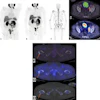
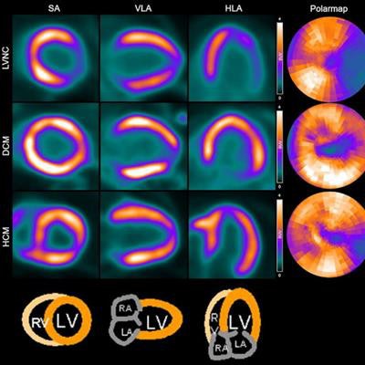
 Example of Ga-68 FAPI-04 PET images in representative patients. LVNC = Left ventricular noncompaction; DCM = Dilated cardiomyopathy; HCM = Hypertrophic cardiomyopathy; SA = Short axis; HLA = Horizontal long axis; VLA = Vertical long axis; RV = Right ventricle; LV = Left ventricle; RA = Right atrium; LA = Left atrium. Image and caption courtesy of EJNMMI Research through CC BY 4.0.
Heterogeneous Ga-68 FAPI-04 uptake was observed in different subtypes of nonischemic cardiomyopathies, according to the findings. In 22 patients, elevated Ga-68 FAPI-04 uptake was seen in the left ventricle, and 10 patients also showed slightly elevated uptake in the right ventricle. In addition, cardiac uptake values were significantly correlated with enlarged ventricular volume evaluated by echocardiography.
"FAPI PET/CT presents a potential value for in vivo visualization and quantification of fibroblast activation on the molecular level," the researchers wrote.
Ultimately, the study was limited by a relatively small sample size and the lack of a control group, yet the authors suggested it provides important preliminary data on the potential use of the FAPI-PET signal as a noninvasive biomarker for non-ischemic cardiomyopathies.
"Further study is warranted for investigating the theranostic and prognostic value of elevated FAP signal," the group concluded.