Ga-68 HER2-PET/CT scans can effectively predict which HER2-positive breast cancer patients will achieve complete response to neoadjuvant therapy, with the ability to identify therapy-sensitive patients early enough to allow treatment adjustments before surgery.
- 70% of patients in the study achieved pathologic complete response to neoadjuvant therapy, while 30% showed non-response
- Ga-68 HER2-PET/CT scans performed after cycle 2 of treatment identified therapy-sensitive patients with 85% positive predictive value for complete response
- An optimal cutoff of SUVmax ≤1.15 at PET2 provided a 15% higher absolute likelihood of complete response compared to nonsensitive subgroups
- This molecular imaging technique overcomes limitations of existing tools like MRI, which struggles to differentiate residual tumor from treatment-induced fibrosis
- Early identification of nonresponders enables timely treatment modifications before surgery, improving patient outcomes
PET/CT scans can identify which patients with HER2-positive breast cancer will achieve complete responses to neoadjuvant therapy, according to a recent study.
A group from the Fourth Hospital of Hebei Medical University and Hebei Provincial Tumor Hospital in Shijiazhuang, China, performed serial PET/CT scans in 45 patients with newly diagnosed disease during treatment, with findings suggesting the modality can flag nonresponders early enough to allow timely changes, noted lead author Sinuo Gao, MD, and colleagues.
“With neoadjuvant therapy’s growing use in the treatment of breast cancer, noninvasive, reproducible, and targeted strategies for predicting and monitoring therapeutic response are increasingly emphasized,” the group noted. The study was published August 6 in the Journal of Nuclear Medicine.
Monitoring neoadjuvant therapy response in HER2-positive breast cancer is challenging with existing tools, the authors wrote. MRI is guideline-recommended, yet struggles to differentiate residual tumor from treatment-induced fibrosis, while F-18 FDG-PET/CT is prone to false positives and fails to capture the molecular heterogeneity of HER2-positive disease.
Alternatively, Ga-68 HER2-PET/CT has emerged as a promising molecular imaging technique for visualizing the expression of the HER2 protein on cancer cells, according to the authors. Despite its advantages, evidence regarding its use in assessing the efficacy of neoadjuvant therapy for breast cancer remains scarce, they wrote.
To test whether Ga-68 HER2-PET/CT could meet the need, the researchers enrolled 44 patients with newly diagnosed stage II-IIIC HER2-positive breast cancer between June 2023 and August 2024. All patients received at least four cycles of trastuzumab combined with pertuzumab or pyrotinib-based neoadjuvant therapy, followed by surgery. Ga-68 HER2-PET/CT scans were performed at baseline (PET1), after cycle 2 (PET2), and before surgery (PET3). The primary endpoint was predictive value for pathologic complete response.
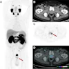
 Ga-68 HER2-PET/CT images before neoadjuvant therapy, at end of second cycle, and before surgery. (A) Baseline scan demonstrated soft-tissue lesion measuring approximately 2.3 × 2.2 cm in upper outer quadrant of left breast, with marked radiotracer uptake and SUVmax of 10.1. (B) At end of second cycle, imaging revealed residual lesion of approximately 2.2 × 1.0 cm in same region, without abnormal radiotracer accumulation. (C) Preoperative imaging demonstrated further reduction in lesion size to approximately 2.0 × 0.6 cm, with no abnormal tracer uptake detected. Journal of Nuclear Medicine
Optimal cutoffs for predicting pCR included an SUVmax of ≤1.15 at PET2 (area under the curve [AUC] = 0.738) and ≤ 0.50 at PET3 (AUC = 0.735). Applying the PET2 threshold identified a therapy-sensitive subgroup with an 85% positive predictive value for pCR, representing a 15% higher absolute likelihood of pCR compared with the nonsensitive subgroup, while the method provided a negative predictive value of approximately 60%, the researchers reported.
“Dynamic changes in 68Ga-HER2 PET/CT uptake during [neoadjuvant therapy] provide an effective early indicator of therapeutic sensitivity in HER2-positive breast cancer,” the group wrote.
While validation in larger, multicenter cohorts is needed before the approach can be broadly adopted, the findings support the value of Ga-68 HER2-PET/CT for predicting pathologic response at both early treatment and preoperative stages, the authors concluded.
Read the full study here.